Figure 2. Illustration of the subretinal
injection technique. A bent needle was inserted through a sclerotomy at
10 o’clock next to the limbus and pushed forward in a tangential
direction toward the retina without touching the crystalline lens (A).
Three to 21 days after inoculation, choroidal neovascularization
membranes (asterisk) could be identified in the subretinal space next
to the optic nerve by light microscopy (B, C). The retinal (C)
and sceral (D) insertion sites are highlighted by arrows. B-D:
Periodic acid-Schiff (PAS) staining was used and original
magnifications are 4X (B) and 20X (C,D).
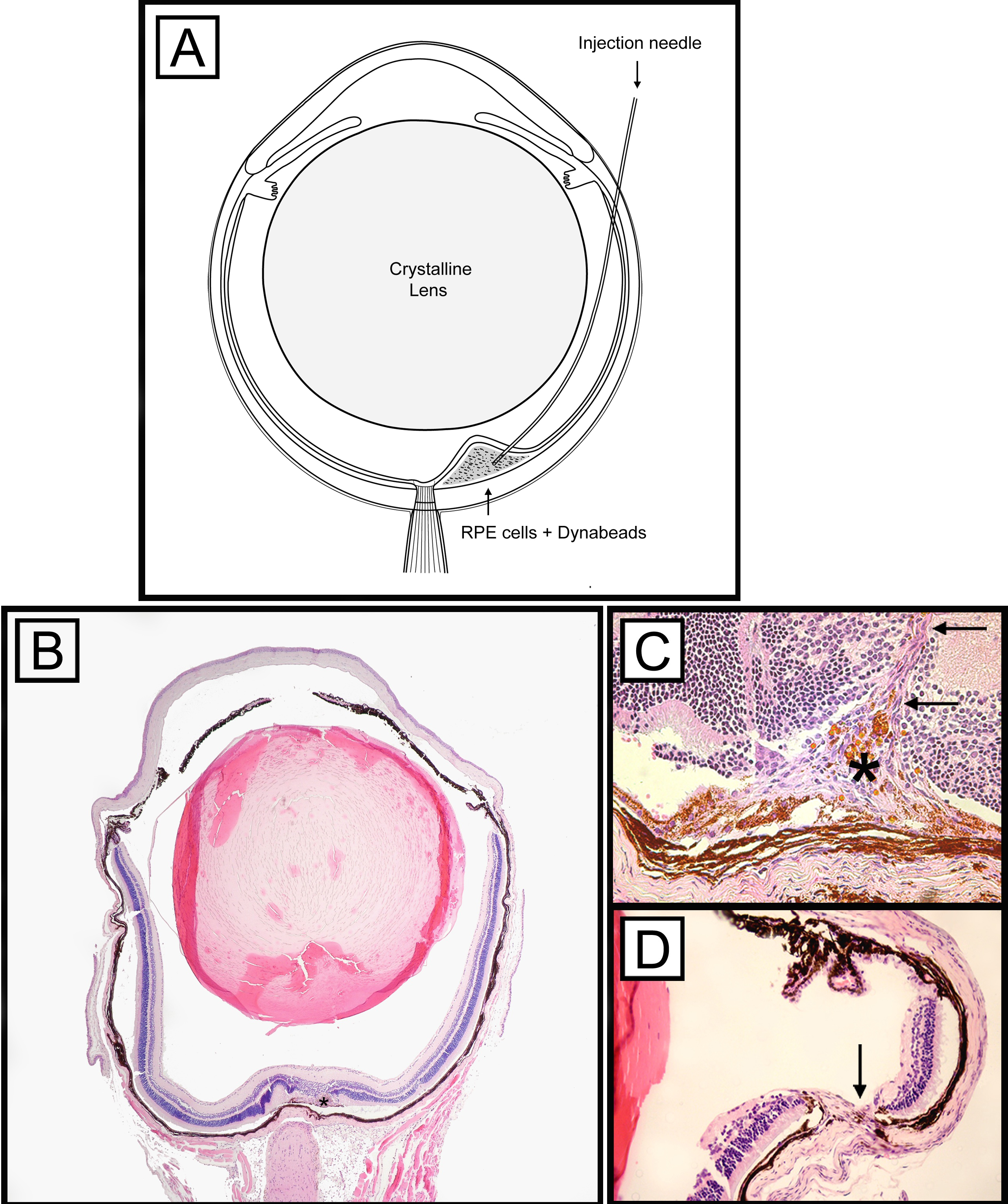
 Figure 2 of Schmack, Mol Vis 2009; 15:146-161.