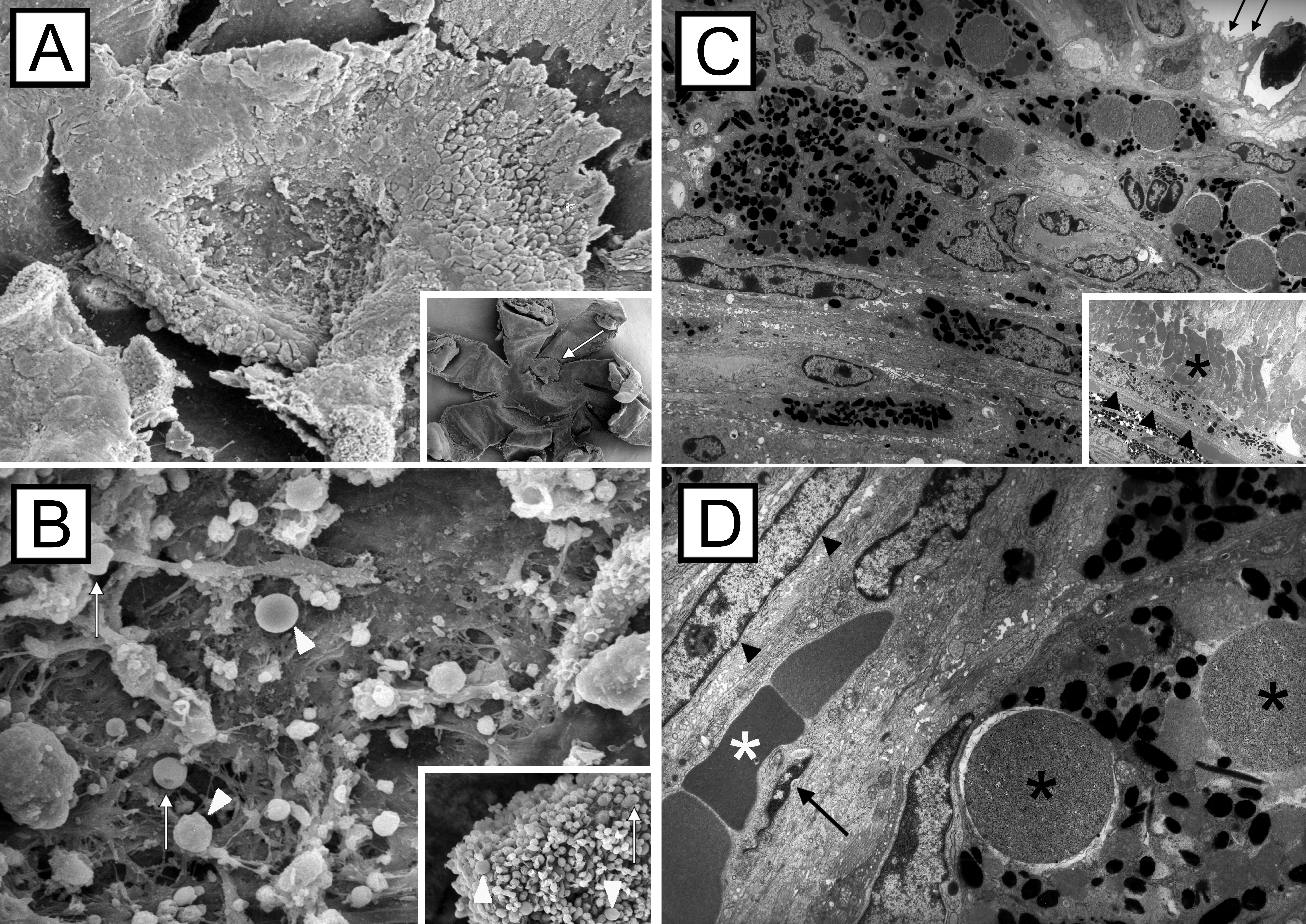
Figure 10. Scanning and transmission
electron microscopic images of choroidal neovascularization membranes.
Choroidal neovascularization (CNV) membranes (A, arrow) were
partially covered by remaining photoreceptor cells after removal of the
retina. Occasional microbeads (B, arrowheads) and
doughnut-shaped erythrocytes are present at the inner CNV surface (B,
arrows). CNV membranes were composed of fibroblasts, pigmented retinal
pigment epithelial (RPE) cells, and non-pigmented RPE cells; a well
structured surface was missing (C, arrows). The RPE monolayer (C,
arrowheads) and photoreceptor outer segments (asterisks) next to the
CNV margin appeared normal. High magnification images of the CNV
lesions revealed pigment-laden RPE cells with intracytoplasmatic
microbeads (black asterisks), spindle-shaped fibroblast (arrowheads),
endothelial cells (arrow), and erythrocytes (white asterisk) within
small-sized blood vessels (D).

 Figure 10 of Schmack, Mol Vis 2009; 15:146-161.
  Figure 10 of Schmack, Mol Vis 2009; 15:146-161.