Figure 6. Immunohistochemistry for IDO and
CD3+ and CD8+ lymphocytes in the corneal endothelium. A-B:
IDO; C: CD3+ T – lymphocytes; D: CD8+ T - lymphocytes.
Magnification A-D is 400X; inlays in A-D
represent twofold enlarged details from respective images marked by
arrows. A: non-rejected cornea negative for IDO (A) and
no T-cell infiltration (data not shown); B-D: rejected
cornea transplant positive for IDO (B) with infiltration of CD3
+ T-lymphocytes (C; arrow, arrowhead) and CD8+ T-lymphocytes (D;
arrow and arrowhead) within IDO-positive (B) cornea. B-D
are images from serial sections.
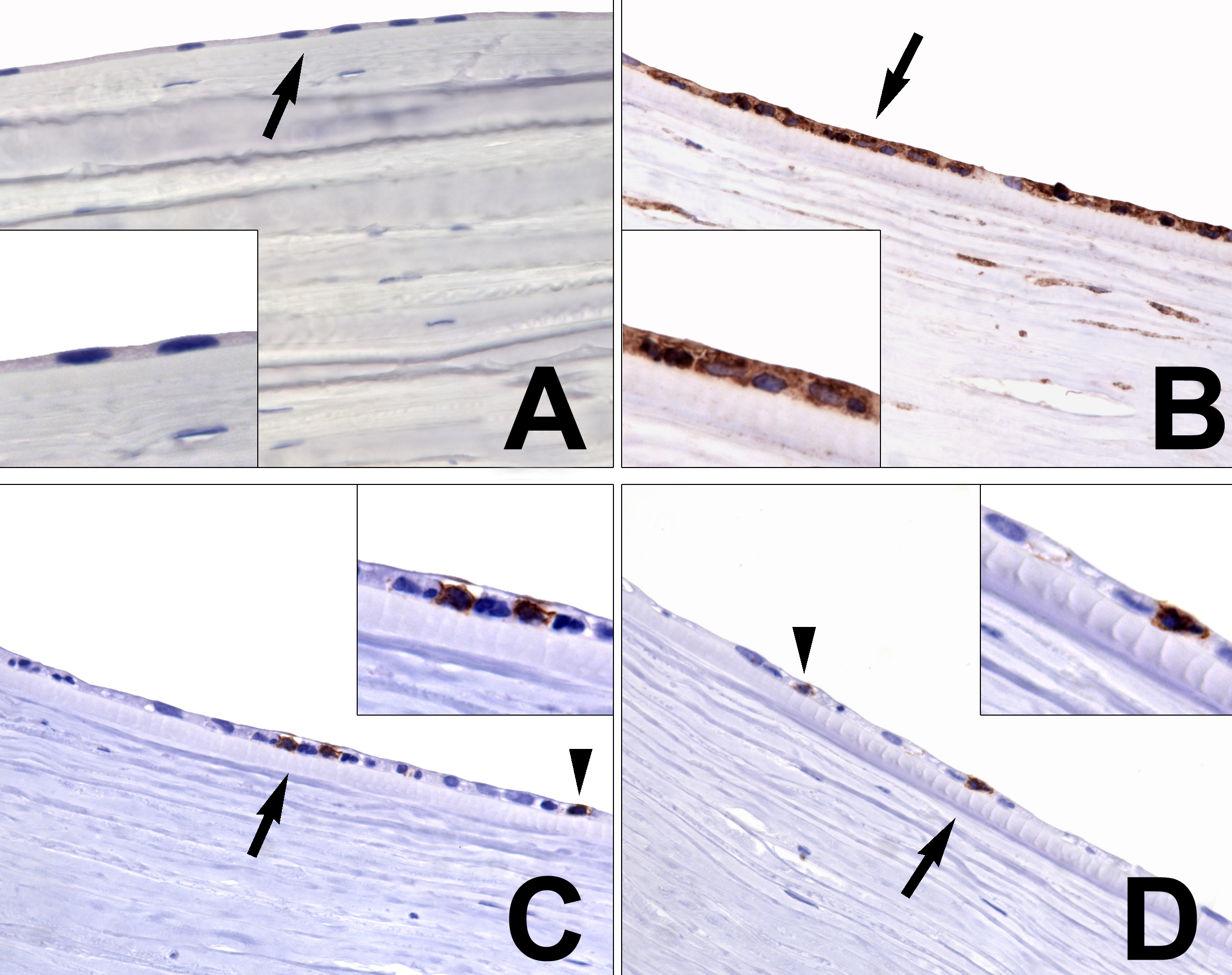
 Figure 6 of Serbecic, Mol Vis 2009; 15:1312-1324.