Figure 1. Clinical progression of fungal keratitis induced by A. fumigatus in mice. The images in the first row (A, B, C, and D) show disease process one, three, five, and seven days, respectively, post-infection in the infected group. The images in
the second row (E, F, G, and H) show the disease process one, three, five, and seven days, respectively, post-infection in the MIP-2 treated group. Here,
the corneal lesion was reduced at all time points. The images in the third row (I, J, K, and L) show the disease process at one, three, five, and seven days, respectively, post-infection in the IL-1β treated group. The
corneal lesion was significantly reduced on day 1 and 3 post-infection.
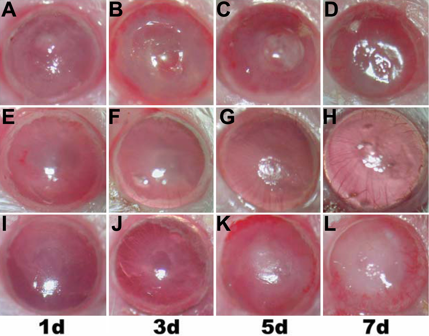
 Figure 1 of
Zhong, Mol Vis 2009; 15:1303-1311.
Figure 1 of
Zhong, Mol Vis 2009; 15:1303-1311.