Figure 5. Immunofluorescent staining of human corneal epithelial culture from limbal explants. A: Staining of K15 in cytoplasm of cultured corneal epithelial cells was observed. B: Cells also showed immunoreactivity for K19 in cytoplasm. C: Expression of ABCG2 in the cell membrane and cytoplasm was seen. D: Some cells revealed positive staining for p63 in nucleus. E: No staining was observed with negative control. Magnification: 400X.
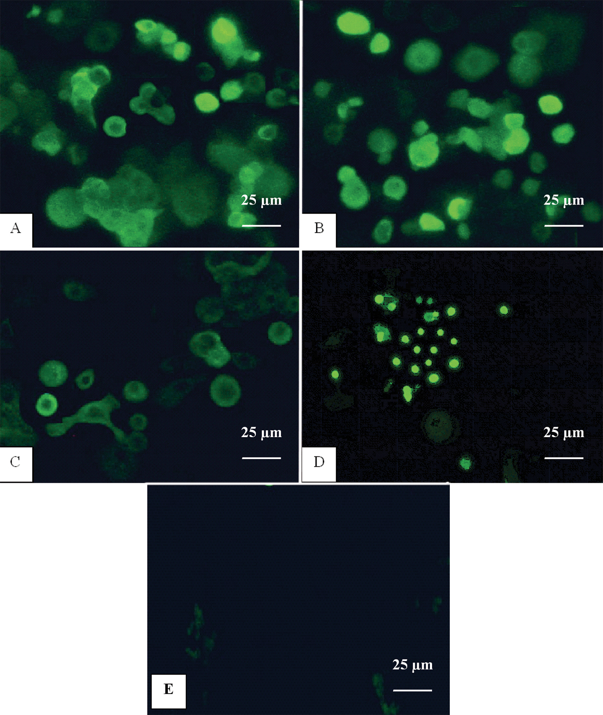
 Figure 5 of
Lekhanont, Mol Vis 2009; 15:1294-1302.
Figure 5 of
Lekhanont, Mol Vis 2009; 15:1294-1302.