Figure 2. Demonstration of limbal cultures
as observed under phase contrast microscope. The corneal epithelial
cells were proliferating from the periphery of the explant (red arrow)
onto the denuded human amniotic membrane in the absence of feeder cells
and serum. The cells adjacent to the explant appeared to be smaller and
more uniform and had large nuclei (white arrow) compared to the cells
that expanded further away from the explant (black arrow).
Magnification: 200X.
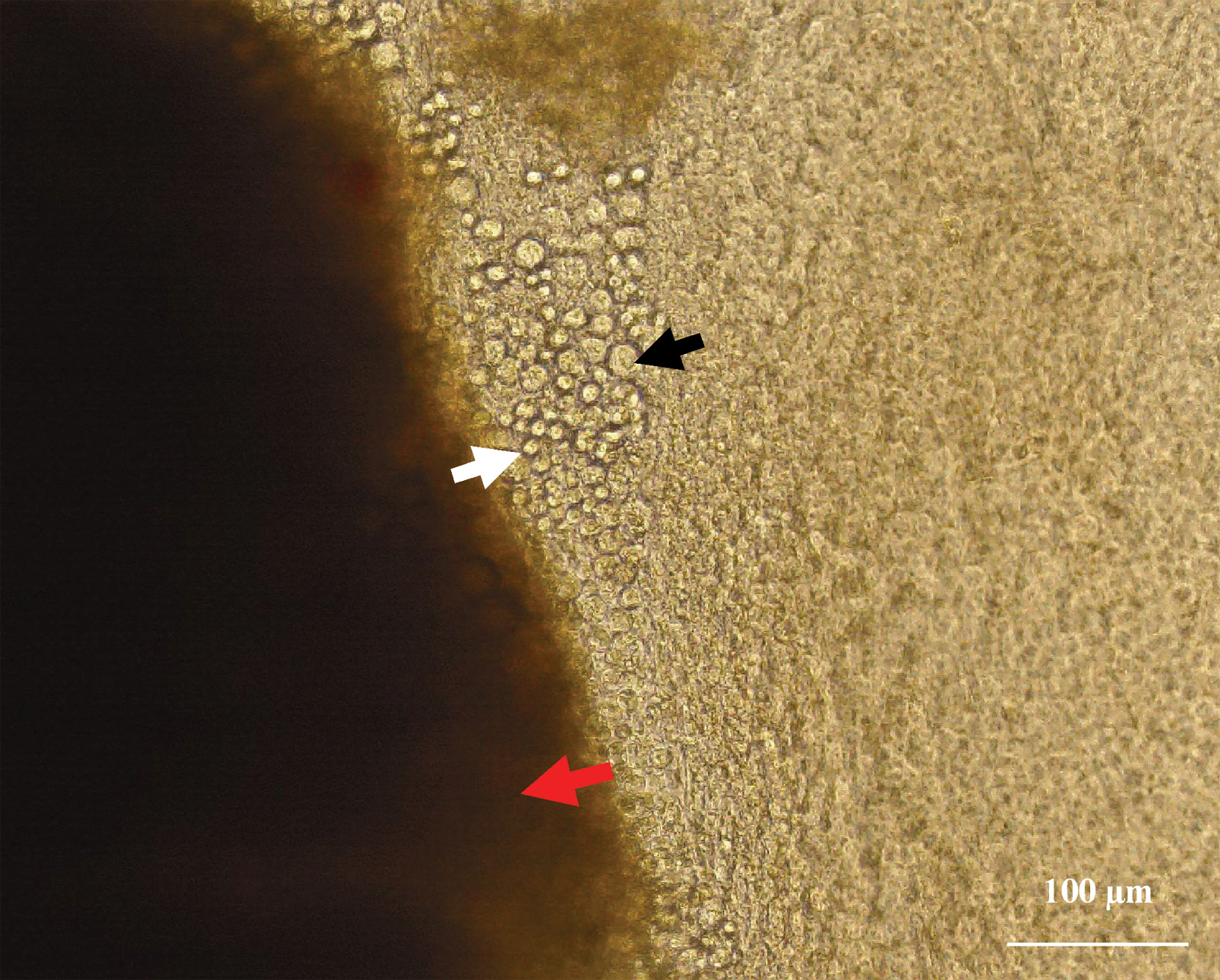
 Figure 2 of Lekhanont, Mol Vis 2009; 15:1294-1302.