Figure 2. Immunohistochemistry of mAChR subtypes from human and Balb/c mouse. A shows the image of the human mAChR subtypes, and B illustrates the image of the Balb/c mouse mAChR subtypes. Subtype selective antibodies bound to scleral fibroblasts (arrowheads)
demonstrates the presence of M1-M5 receptors (shown in green and nucleus shown in blue; stains with DAPI) at magnification 400X. When secondary FITC-labeled
antibody was used without the primary antibody, no significant binding was observed (-ve cont means negative control). Arrow
indicates scleral fibroblast. Scale bar=50 μm. Inset in B: enlarged image of fibroblast. All experiments were done in triplicate.
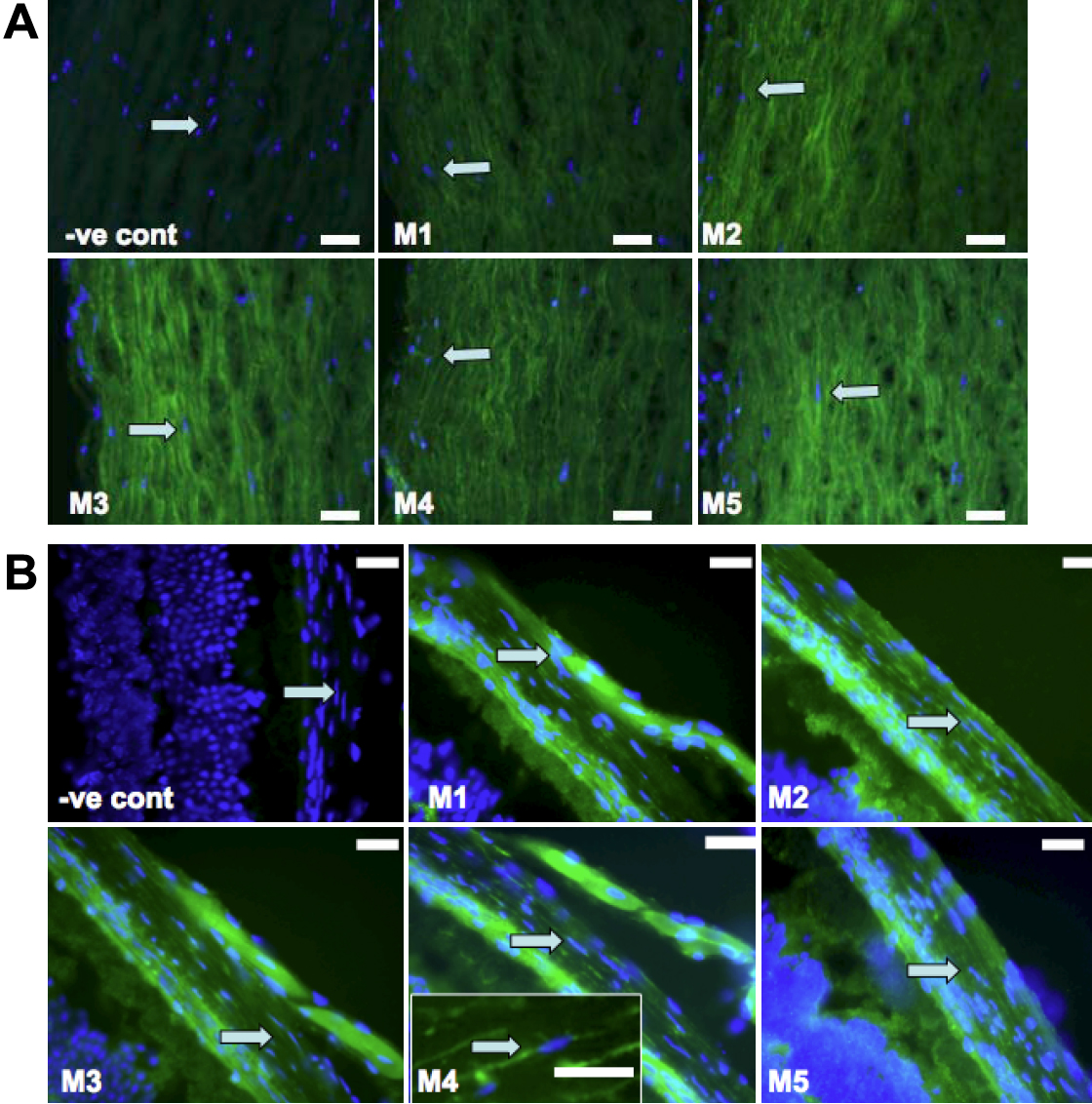
 Figure 2 of
Barathi, Mol Vis 2009; 15:1277-1293.
Figure 2 of
Barathi, Mol Vis 2009; 15:1277-1293.