Figure 3. Photomicrographs of the lens and hyaloid vasculature in eyes from E18.5 mice treated with either vehicle-only, TNP-470, or
AS4.5 solution. Low power micrographs (A-C) and matching high power detail (D-F) of the lens and hyaloid vasculature in eyes from E18.5 mice stained with toluidine blue are displayed. Dams were treated
with either vehicle-only (control: A,D), TNP-470 (B,E), or AS4.5 (C,F). In high power light micrographs (D-F), the hyaloid vessels are clearly visible on the lens surface (arrows) in addition to hyalocytes (arrowheads). Scale bars
on A-C=100 µm and on D-F=50 µm.
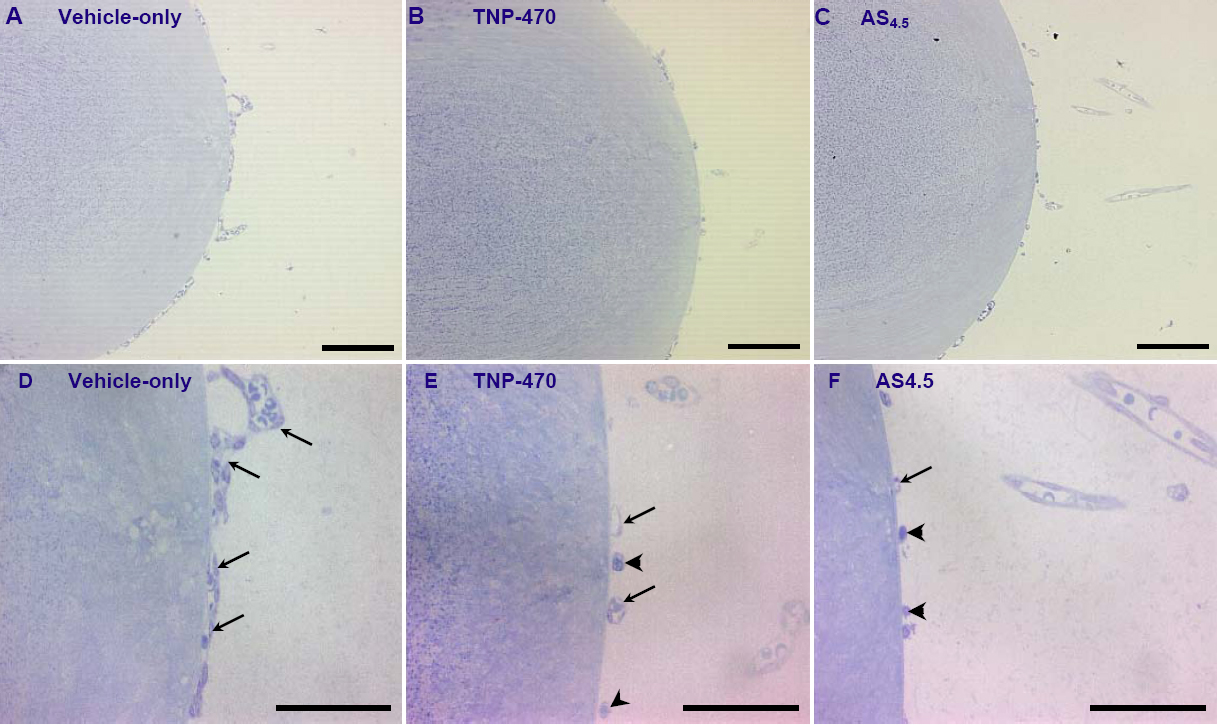
 Figure 3 of
Rutland, Mol Vis 2009; 15:1260-1269.
Figure 3 of
Rutland, Mol Vis 2009; 15:1260-1269.