Figure 4. Influence of LOR on the
histopathological and immunohistochemical studies in mouse corneas 14
days after UV irradiation. The typical histologic findings of cornea
stained with hematoxylin and eosin (A, B) are shown. A:
The cornea in the saline-treated group shows marked inflammation,
obvious edema, profound neovascularization, and significant
hypercellularity in the stroma. B: The cornea in the
LOR-treated group exhibits only scattered inflammatory cells, mild
stromal swelling, and less neovascularization. Corneal tissues (C-F)
were analyzed by immunohistochemistry to determine the expression of
NF-κB. Immunohistochemical staining with an antibody against activated
NF-κB was performed to detect the expression of NF-κB. Sections
incubated without a primary antibody served as negative controls. All
tissue sections were counterstained with hematoxylin. These samples
were representative of all corneas examined. Brown staining indicates
activated NF-κB. C,D: The cornea in the mock-infected
group and the cornea in LOR alone group show that NF-κB activity is
only observed very faintly in the base cells of epithelium
(arrowheads). E: Recurrence induced wide spread positive
staining of NF-κB, which was most robust in the stroma (arrows) of the
saline-treated group. F: Scant immunoreactivity of NF-κB was
observed in the stroma of the LOR-treated group (arrows). Original
magnifications, 400X.
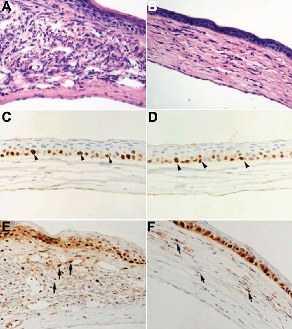
 Figure 4 of Yin, Mol Vis 2009; 15:1252-1259.