Figure 7. The annexin A2 expression in
vascular endothelial cells . At P15, mice reared in room air (A, E, C)
or mice with hypoxia-induced retinopathy (B, D, F) were killed
(n=6), and ocular frozen sections were stained with a rat antibody
directed against CD31 or a rabbit anti-murine annexin A2 antibody (B,
D, F). Secondary antibodies were a goat anti-rabbit IgG conjugated
with FITC and a Cy3-conjugated donkey anti-rat antibody. Small
cross-sections of retinal vessels are evident in the CD31-stained
retina (red) from a normal mouse, while dilated vessels and new vessels
can be seen in the CD31-stained retina from a mouse with ischemic
retinopathy. There is no detectable staining for annexin A2 in the
retina of the mouse reared in room air, but there is prominent staining
in the retina of the mouse with ischemic retinopathy (green). Merging
of the images for the retina of the mouse with ischemic retinopathy
(bottom, right) by simultaneously viewing with the red and green
channels, demonstrates colocalization of CD31 and annexin A2,
indicating that annexin A2 is expressed in vascular endothelial cells.
Scale bar represents 100 μm.
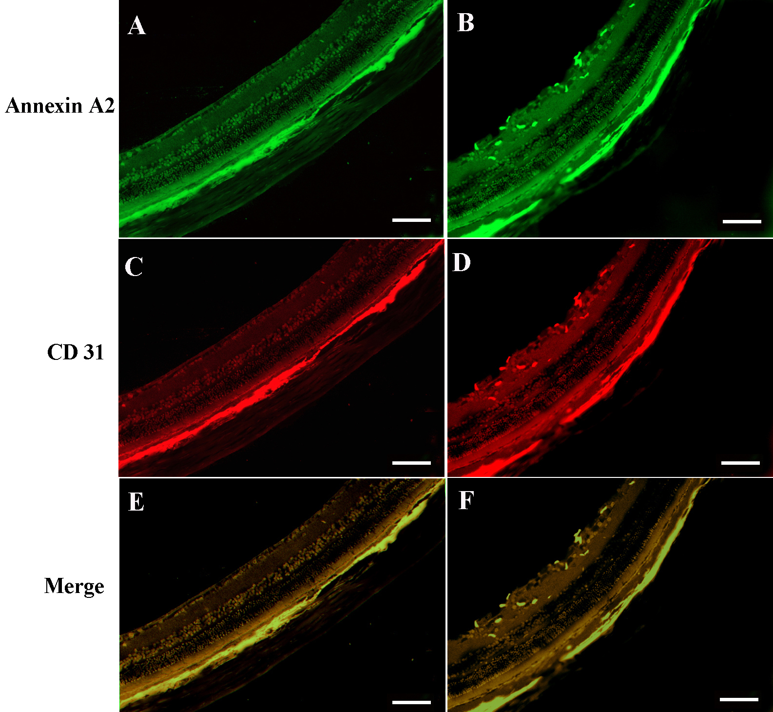
 Figure 7 of Zhao, Mol Vis 2009; 15:1231-1242.