Figure 6. OCT3 scans (6 mm) centered on the fovea. From the right eye (A) and left eye (B) of affected individual III:4. Hyperreflectivity (*) at the level of the RPE-choroid complex is noted throughout the macula
in the right eye (OD) and to a lesser extent in the left eye (OS). The right eye also demonstrates intraretinal cysts throughout
the macula and a large central retinal detachment. The left eye demonstrates flattening of the foveal pit and loss of the
outer nuclear layer temporal to the fovea.
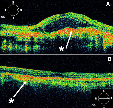
 Figure 6 of
Saihan, Mol Vis 2009; 15:1218-1230.
Figure 6 of
Saihan, Mol Vis 2009; 15:1218-1230.