Figure 5. OCT3 scans (6 mm) centered on the fovea. From the right eye (A) and left eye (B and C) of affected individual III:3. The right eye (OD) demonstrates inframacular retinal thickening due to intraretinal cystic
changes (IRC). The left eye (OS) demonstrates central retinal thinning and both retinal and pigment epithelial detachment
(RPED) superior to the fovea. In the left eye there is hypereflectivity at the level of the choroid nasal to the fovea.
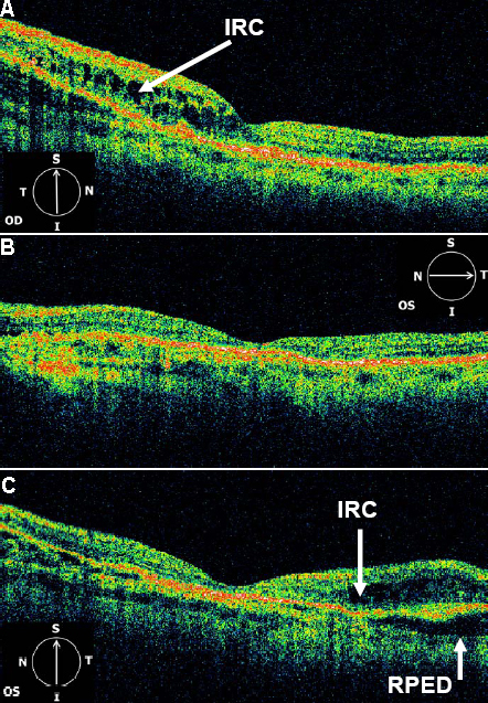
 Figure 5 of
Saihan, Mol Vis 2009; 15:1218-1230.
Figure 5 of
Saihan, Mol Vis 2009; 15:1218-1230.