Figure 4. OCT3 scans (6 mm) centered on the fovea. (A) from a normal adult individual. Landmarks such as outer nuclear layer (ONL), inner and outer photoreceptor segment junction
(IS), retinal pigment epithelium (RPE), and choriocapillaris (CC) are indicated. (B) from affected individual III:1 Right eye (OD) C: Individual III:1 Left eye (OS). Both B and C demonstrate hyperreflective signals (*) at the level of the RPE-choroid complex. The foveal pit appears steep in the right
eye (OD). Parafoveal intraretinal cysts (IRC) are demonstrated bilaterally. There is also evidence of parafoveal retinal atrophy
in the left eye (OS).
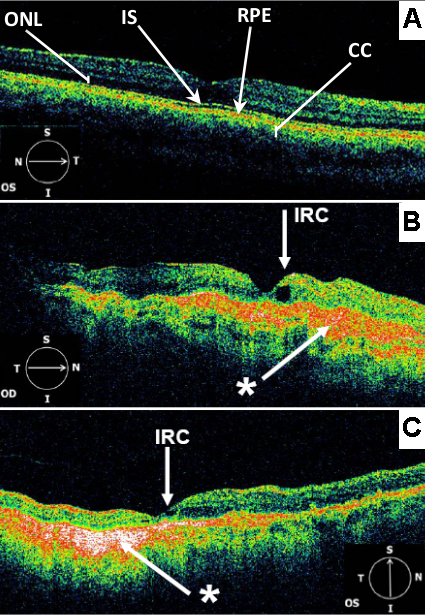
 Figure 4 of
Saihan, Mol Vis 2009; 15:1218-1230.
Figure 4 of
Saihan, Mol Vis 2009; 15:1218-1230.