Figure 3. Color fundus photographs of the three affected individuals. All three affected individuals demonstrated the presence of yellow
drusen-like deposits bilaterally throughout their fundi, but to varying amounts. A: Individual III:1 had bilateral geographic
atrophy and widespread subretinal drusen-like deposits throughout the posterior pole extending beyond the vascular arcades.
B: Individual III:3 had subretinal and intraretinal fluid at the macula in the right eye extending inferiorly toward the temporal
arcades. In the right eye, two extrafoveal foci of subretinal hemorrhage were also noted, inferior to the optic disc and temporal
to the macula. In the left eye (poorest visual acuity), subretinal fluid and a pigment epithelial detachment were noted superior
to the macula. Although not visible on this image, areas of chorioretinal atrophy were also noted nasal to the disc. C: Individual
III:4 had a large subretinal fibrovascular scar visible in the right eye. Foci of subretinal hemorrhage, exudates, and secondary
retinal detachment are also visible. Areas of floccular yellowish subretinal deposit can be seen throughout the fundus in
the right eye. In the left eye (without visual loss) only small yellowish subretinal deposits can be seen throughout the posterior
pole.
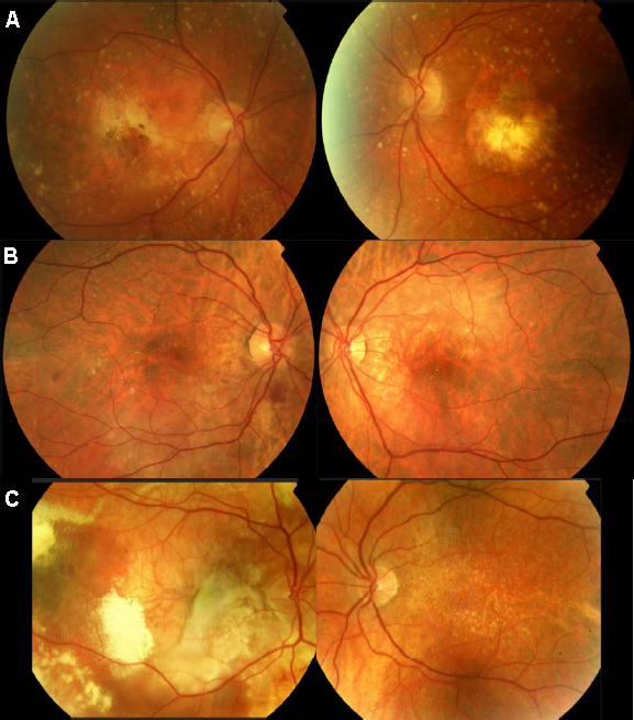
 Figure 3 of
Saihan, Mol Vis 2009; 15:1218-1230.
Figure 3 of
Saihan, Mol Vis 2009; 15:1218-1230.