Figure 2. Electropherogram from affected family member III:4. Representative electropherogram from the sense strand of genomic DNA showing
(A) heterozygous G>A missense change, (B) wild type sequence. The wild-type codon frame and amino acid number (139) is also shown.
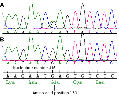
 Figure 2 of
Saihan, Mol Vis 2009; 15:1218-1230.
Figure 2 of
Saihan, Mol Vis 2009; 15:1218-1230.