Figure 1. Photographs of cells analyzed by
comet assay analysis. A: Photograph A depicts intact
cells (without tail) of a patient treated with ranimizumab. B:
Photograph B depicts cells of a patient 30 min after treatment
with PDT. Arrowhead points to a typical “comet” with a bright head and
tail.
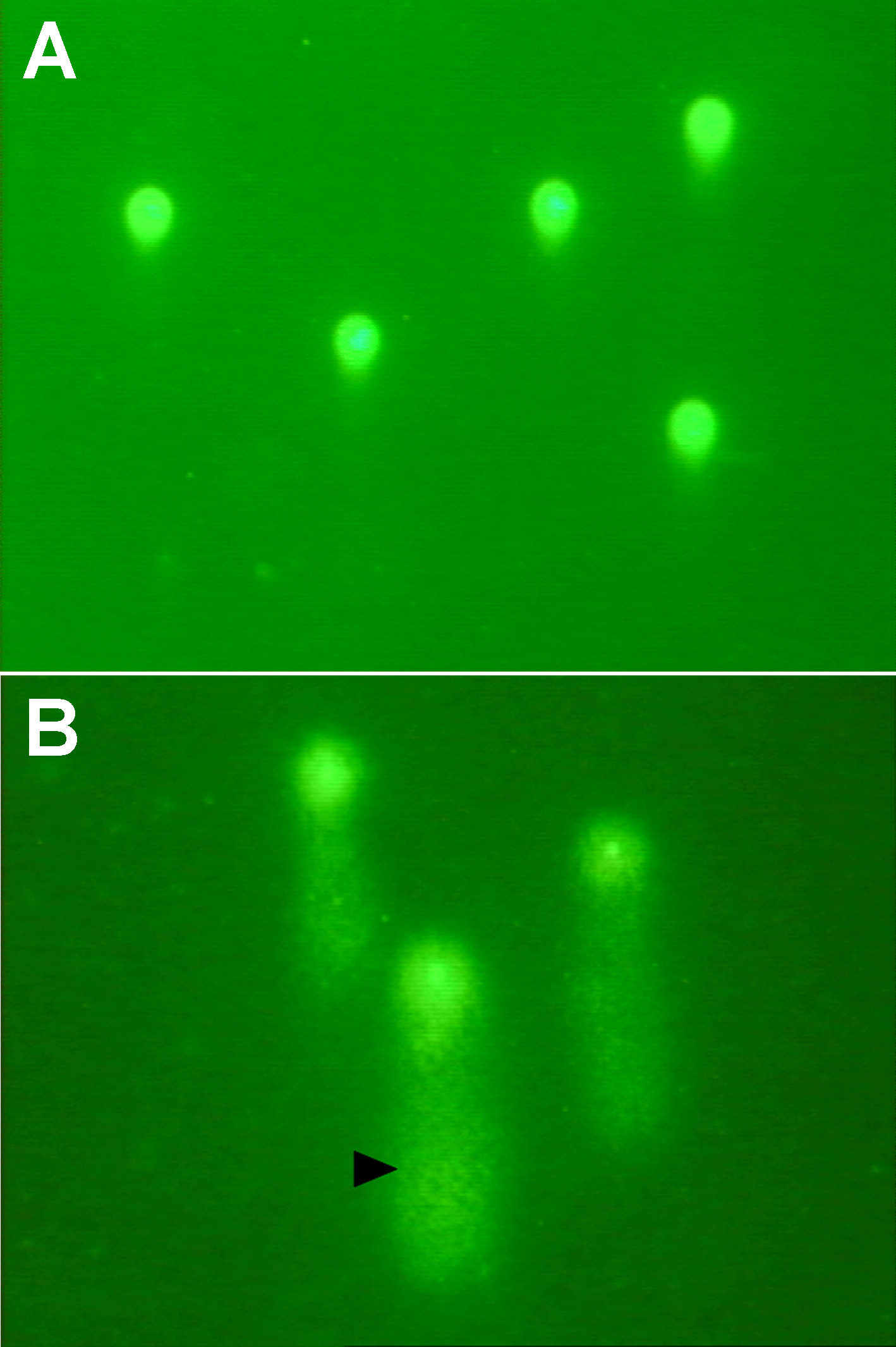
 Figure 1 of Mozaffarieh, Mol Vis 2009; 15:1194-1199.