Figure 2. Representative different calcium
responses of HCECs to PAF. PAF (10 nM) was added at 0 min. The y-axis
shows the intensity ratio of fluorescence at the two excitation
wavelengths of a single cell. High ratio indicates higher [Ca2+]i.
A depicts a minimal response of a cell to PAF. B shows a
rapid response to PAF followed by a relatively rapid decay of the [Ca2+]i
mobilization response. C displays a PAF-induced [Ca2+]i
mobilization response that lasts for at least 16 min. D shows
an apparent oscillatory [Ca2+]i mobilization
pattern in an HCEC. The basal control effects with just the vehicle
resembled the lack of response in A.
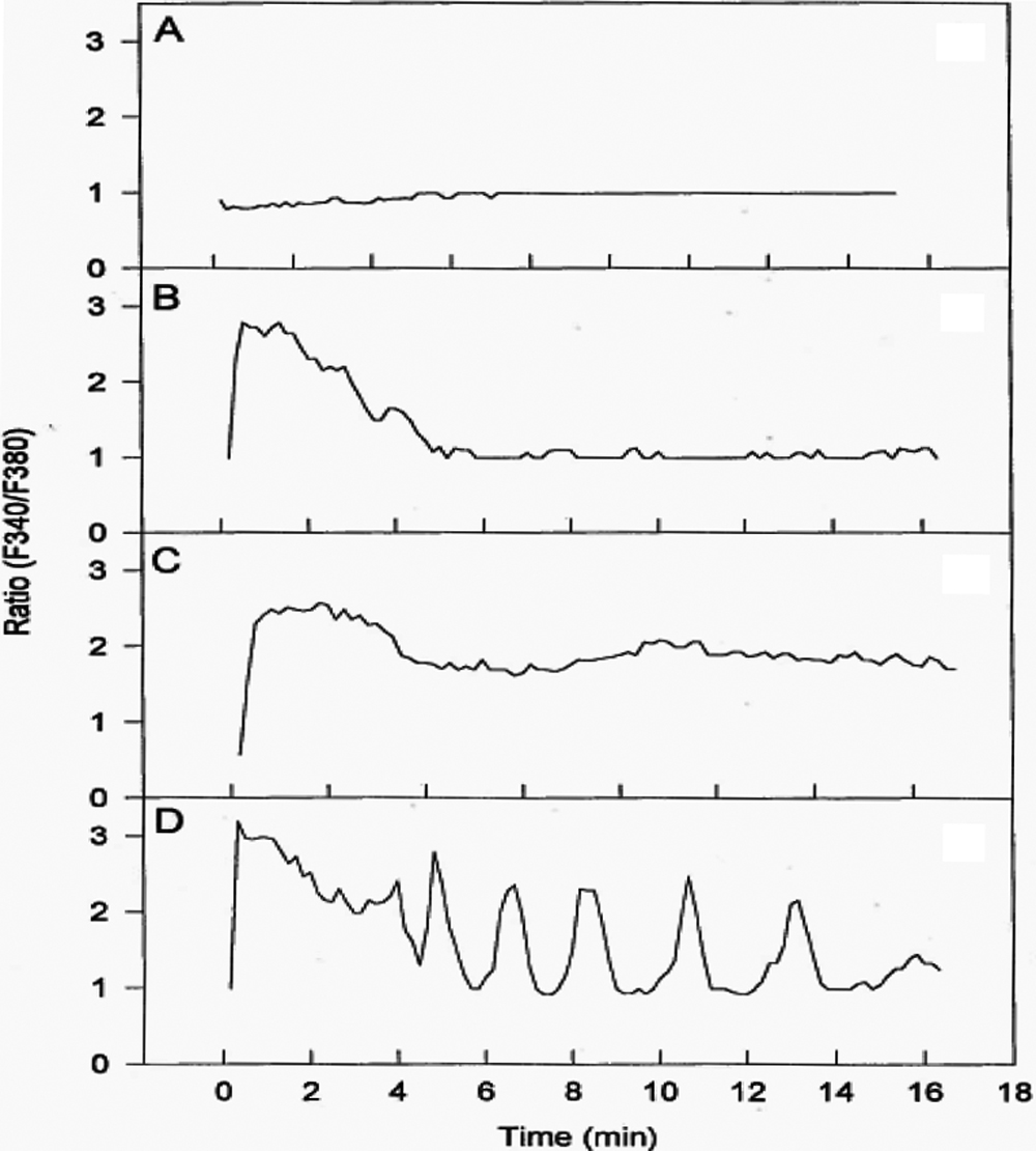
 Figure 2 of Sharif, Mol Vis 2009; 15:1153-1161.