Figure 6. Expression of BER mRNA and
proteins in the adult mouse retina. A: Semiquantitative RT–PCR
experiments were performed to determine the APE1, DNA
polymerase β (DNA pol β), and XRCC1 mRNA levels of
expression in C57BL/6 mouse neuroretinal cells. Cyclophilin A (Cyclo)
was used as an internal control. Specific primers for amplifying mouse APE1,
DNA pol β, XRCC1, and Cyclophilin A cDNAs were used. The expected
size for each specific amplified product was obtained: 446 bp for APE1,
693 bp for DNA pol β, 418 bp for XRCC1, 311 bp for Cyclophilin
A. Immunohistological localization of APE1, DNA polymerase β, and
XRCC1 in the adult mouse retina was performed using an antibody raised
against APE1 (B), DNA pol β (C) or XRCC1 (D). The
staining appears in brown. Sections were counterstained with a methyl
green solution. No signal was detected when the specific anti-APE1
antibody was omitted (E). APE1 and DNA polymerase β were
detected in the ganglion cell layer (GCL), the inner nuclear layer
(INL), and the photoreceptor inner segments (IS). Labeling was also
observed in the INL and outer plexiform layers (ONL). Surprisingly,
XRCC1 was not detected in the IS. Scale bar equals 50 μm in B, C,
and E, and 10 µm in D.
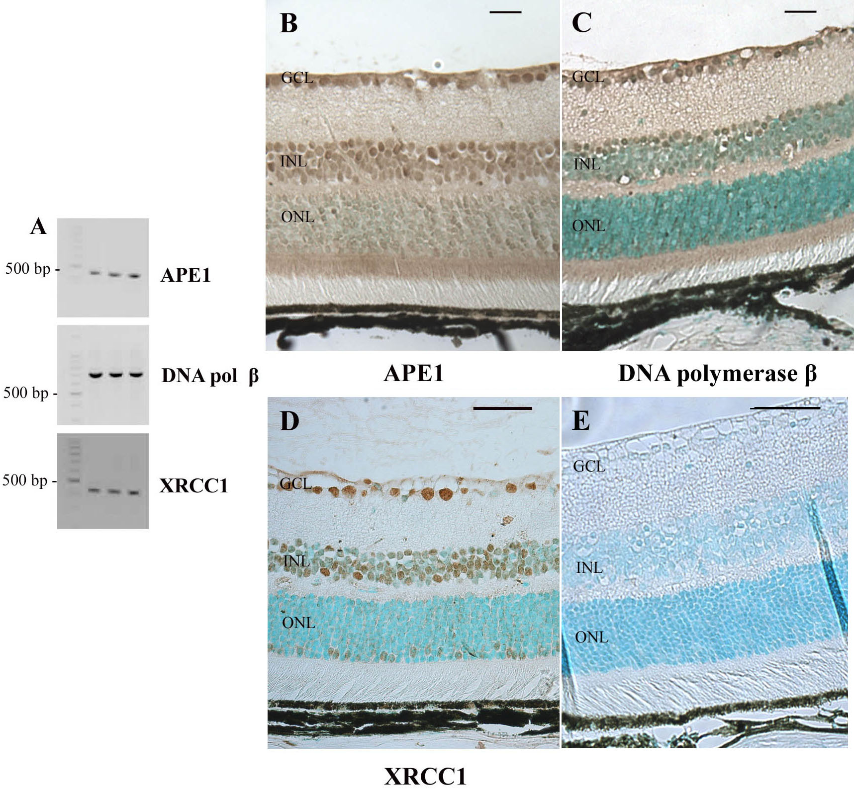
 Figure 6 of Bigot, Mol Vis 2009; 15:1139-1152.