Figure 5. Cellular distribution of Ogg1
protein in the retinal cells. Using an antibody raised against human
8-oxoguanine DNA glycosylase, immunostaining is detected in
photoreceptor inner segments (A), outer nuclear layer (ONL, B),
inner nuclear layer (INL, C) and ganglion cell layer (GCL, D).
Ogg1 immunoreactivity was detected both in the cytoplasmic and nuclear
compartments of the immunolabeled cells of GCL and INL. The staining
seemed only cytoplasmic in photoreceptor cells, which also displayed a
strong labeling in the inner segments (IS). Scale bar equals 20 μm in A,
and 8 µm in B, C, and D. Abbreviations:, inner
plexiform layer (IPL); outer segment (OS); outer plexiform layer (OPL);
retinal pigment epithelium (RPE).
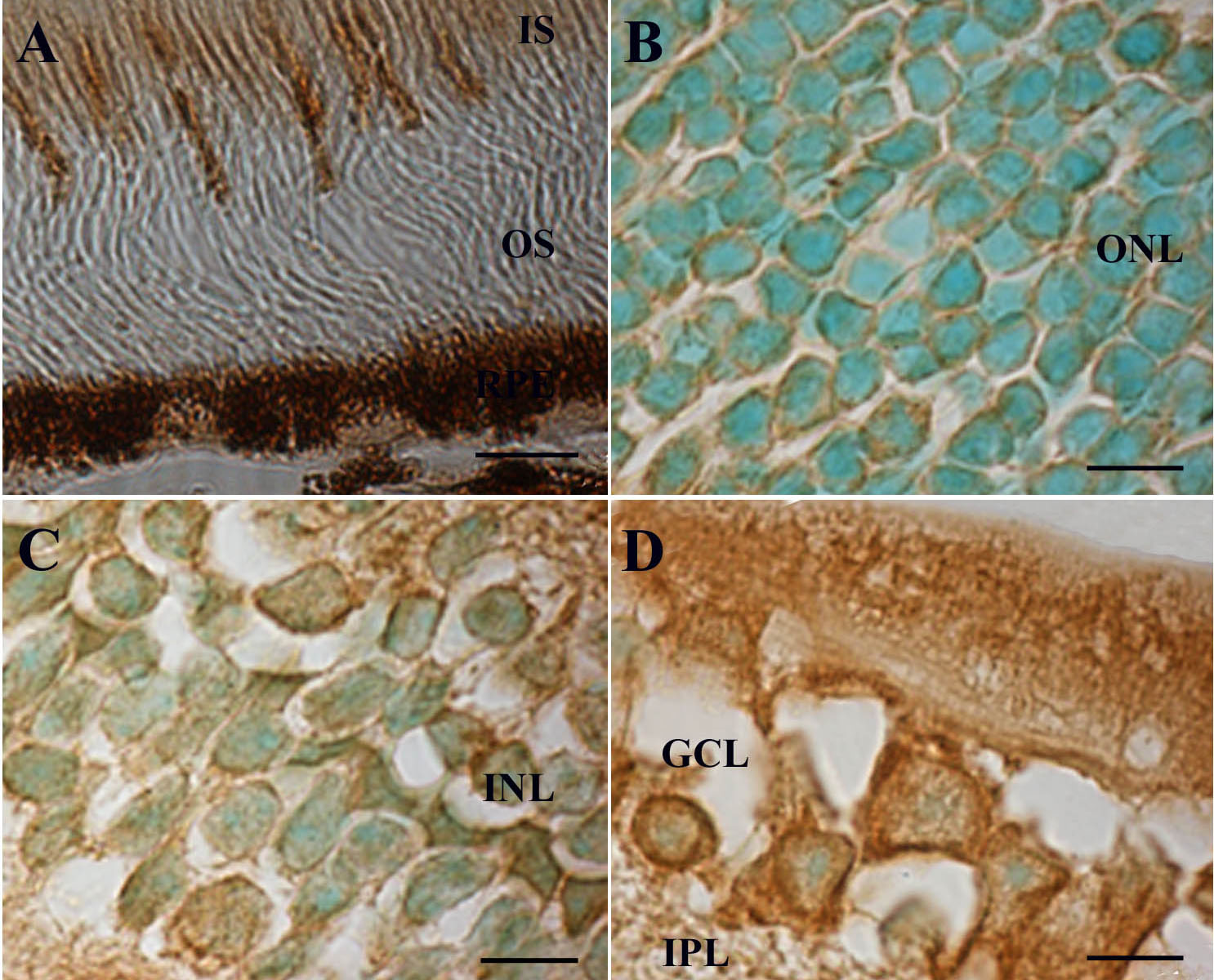
 Figure 5 of Bigot, Mol Vis 2009; 15:1139-1152.