Figure 4. Immunohistochemical localization
of Ogg1 in the adult mouse eye. Using an antibody raised against human
8-oxoguanine DNA glycosylase, immunostaining is detected in in
neuroretina (A),ciliary body (D), cornea (E), and
lens (F). The Ogg1 staining appears in brown, and sections were
counterstained with methyl green solution. No signal was detected when
the specific anti-Ogg1 antibody was omitted (G). Ogg1 protein
was mainly present in the ganglion cell layer (GCL), in photoreceptor
inner segments (IS), and in the outer (OPL) and inner plexiform layers
(IPL). B and C show a high magnification of the inner
and the outer portions of retina, respectively. Ogg1 protein was also
detected in non-neuronal cells: corneal epithelium (Cep), pigmentary
epithelium (PCE) and nonpigmentary ciliary epithelium (NPCE), lens
epithelium (Le), lens transition zone (Tz), and lens fibers (Lf). Scale
bar equals 50 μm in A, D,and G, 20 μm in B, C,
and E, and 45 µm in F. Abbreviations: corneal stroma
(Cs); inner nuclear layer (INL); outer nuclear layer (ONL); retinal
pigment epithelium (RPE).
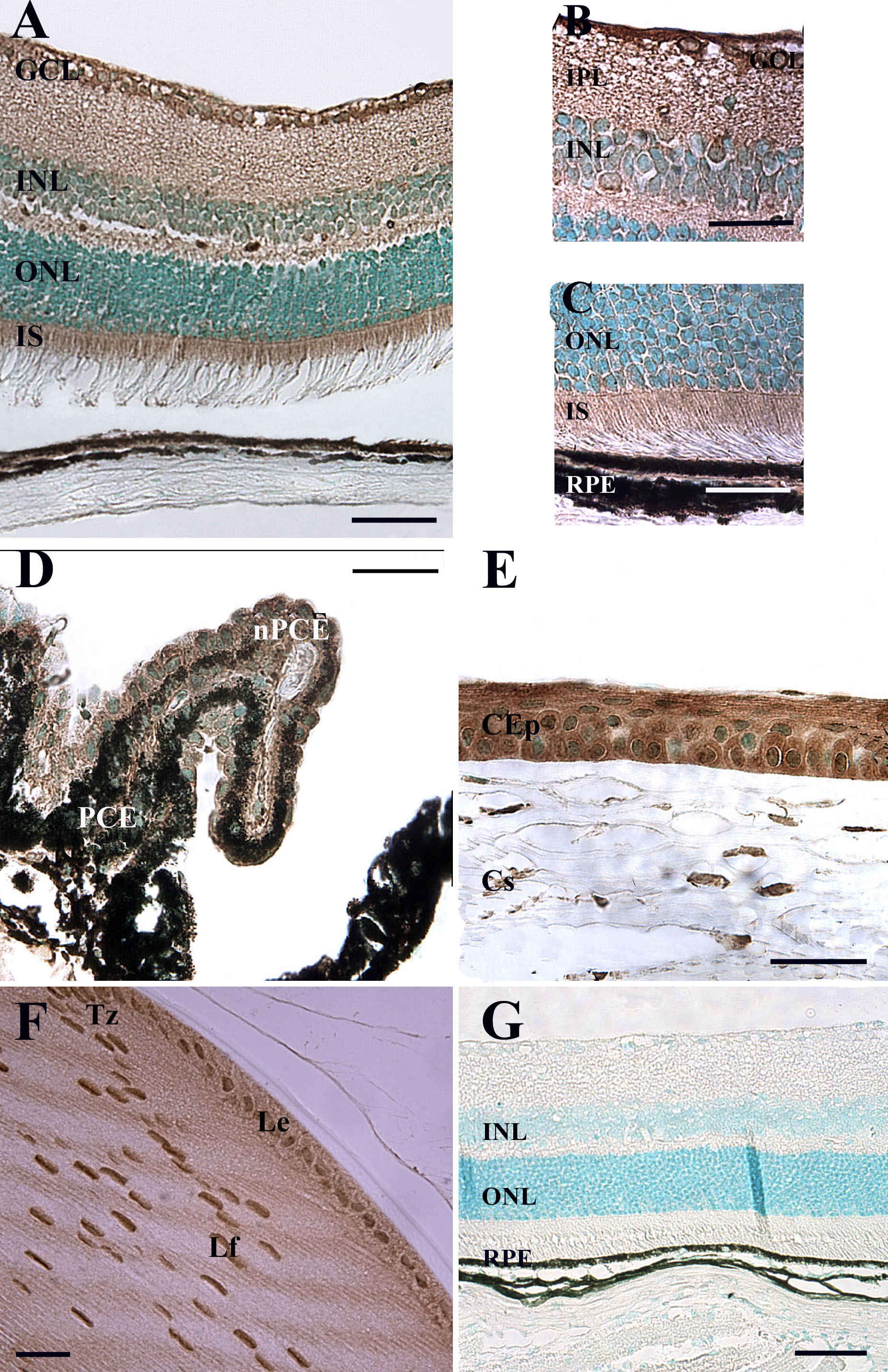
 Figure 4 of Bigot, Mol Vis 2009; 15:1139-1152.