Figure 3. Distribution of Ogg1
transcript in the mouse adult eye. In situ hybridization signals of
8-oxoguanine glycosylase (Ogg1) mRNA were detected in
neuroretina (A, C, and D), ciliary body (E), and
cornea (F) using a specific antisense digoxigenin-labeled
riboprobe. Labeled Ogg1 mRNA appears as purple precipitates. No
signal was detected with a control sense probe (B). Ogg1
mRNA was present in all retinal nuclear layers, in photoreceptor inner
segments (IS), and in retinal pigment epithelium (RPE). Ogg1
mRNA was also detected in non-neuronal cells: corneal epithelial (Cep)
and endothelial cells (Cep), keratocytes of the corneal stroma,
pigmentary epithelium (PCE), and nonpigmentary ciliary epithelium
(NPCE). C and D represent a high magnification of the
inner portion of the neuroretina, the retinal pigment epithelium (RPE),
and the choroid (Ch), respectively. Scale bar equals 70 μm in A, B,
E, and F, and 25 µm in C and D.
Abbreviations: ganglion cell layer (GCL); inner nuclear layer (INL);
outer nuclear layer (ONL).
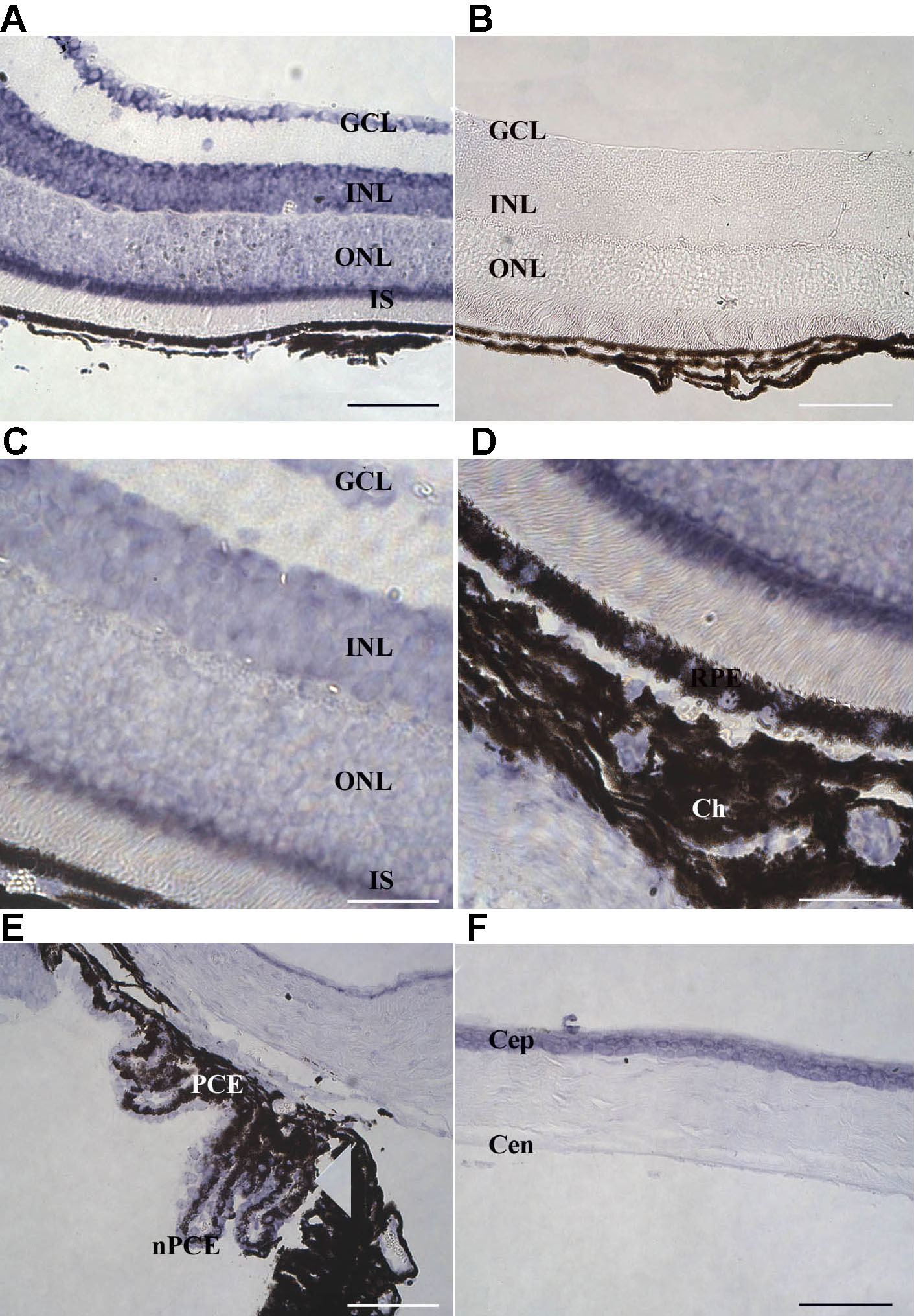
 Figure 3 of Bigot, Mol Vis 2009; 15:1139-1152.