Figure 1. Immunohistological detection of
8-oxoguanine in the mouse retina. Immunostaining using an antibody
raised against 8-oxoguanine (8-oxoG) was detected in liver of wild-type
(A) and Ogg1-deficient mice (B), and in C57BL/6J mouse
retina (C). A stronger signal was detected in liver of
Ogg1-deficient mice as compared to wild-type mice. This confirmed the
accumulation of the modified base. 8-oxoG was present in all retinal
nuclear layers, in photoreceptor inner segments (IS), and in retinal
pigment epithelium (RPE; arrow). The 8-oxoG labeling appears in brown.
No signal was detected when the specific anti-8-oxoG antibody was
omitted (D). Scale bar equals 50 µm in A and B,
25 μm in C, and 60 µm in D. Abbreviations: ganglion
cell layer (GCL); inner nuclear layer (INL); outer nuclear layer (ONL).
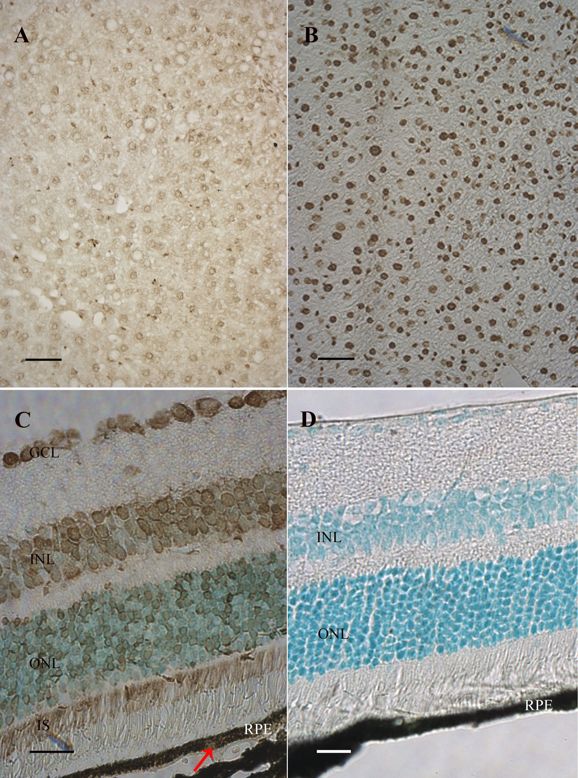
 Figure 1 of Bigot, Mol Vis 2009; 15:1139-1152.