Figure 5. Apoptotic cell death following exposure to elevated hydrostatic pressure. Differentiated RGC-5 cells were exposed to elevated
hydrostatic pressure (30 mmHg) for 3 days. Non-pressurized control cells show normal nuclei (A). However, the apoptotic phenotype of nuclei in RGC-5 cells exposed to elevated hydrostatic pressure was observed (arrows;
B). Size bar represents 20 µm (A and B). Normal and apoptotic nuclei of RGC-5 cells were stained with Hoechst 33342 and counted in several fields under the fluorescence
microscope (for ultraviolet excitation). Fifteen thousand cells in control and pressurized cells were counted, respectively,
and shown as a percentage of cells with apoptotic nuclei among the total cells counted. Data represent the means±SD of three
independent experiments (C). Caspase-3 activation was analyzed by western blotting. The presence of caspase-3 activation was assessed by the observation
of 17 kDa subunit that derived from the cleavage of the 32 kDa pro-enzyme caspase-3 (D). The blot was stripped and reprobed with anti-actin antibody (~42 kDa) to confirm similar protein loading in each lane.
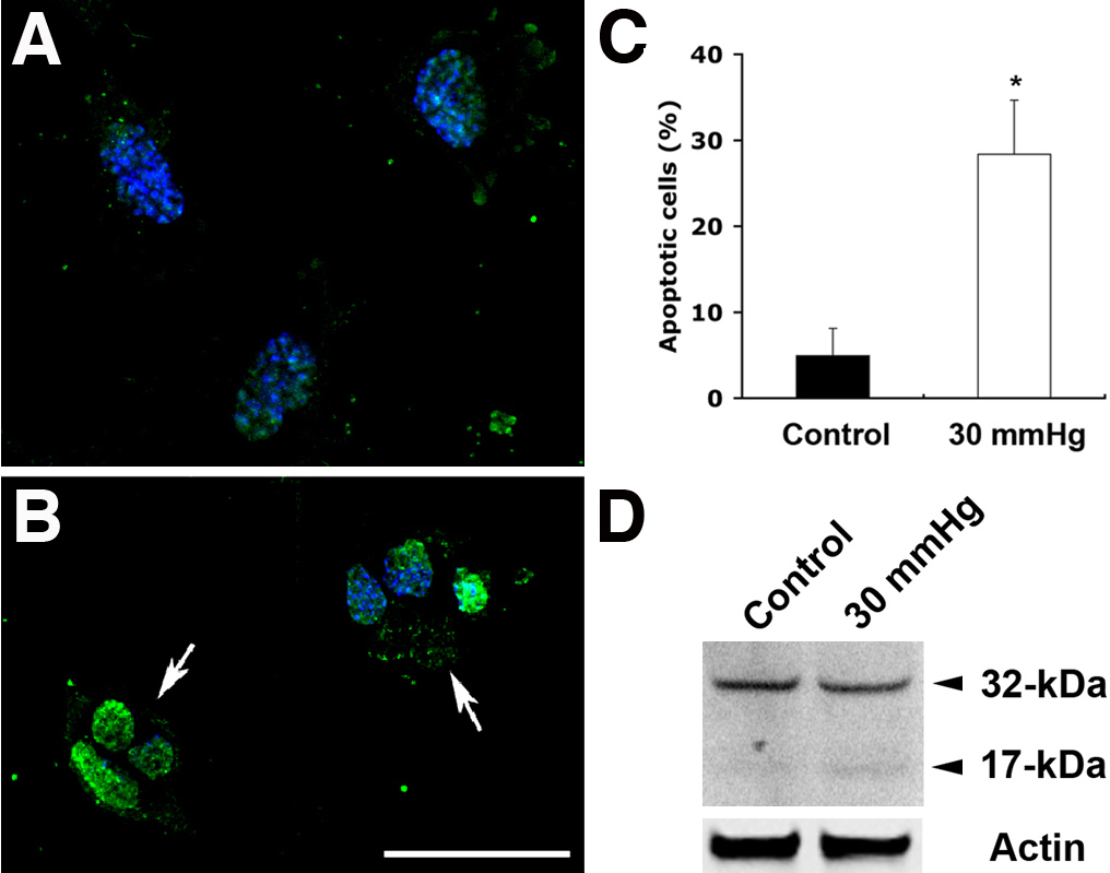
 Figure 5 of
Ju, Mol Vis 2009; 15:120-134.
Figure 5 of
Ju, Mol Vis 2009; 15:120-134.