Figure 5. Western blot analyses of protein
extracts from astrocytes. A representative figure from three repeating
experiments is shown. A: Analysis is shown of protein extracts
from rat brain cortex astrocytes that were subjected to different
durations of pressure. Control cells were allowed to remain in the
incubator at 37 °C without being exposed to any increase in
pressure. Other astrocytes were subjected to an increase in pressure.
Cells were subjected to fixed pressure and then analyzed for iso[4]LGE2–protein
modification. About 3,000 isolated rat brain cortex astrocytes were
subjected to a pressure of 100 mmHg for different time periods
(ranging from 3–33 h). Following pressure treatment, the culture medium
was immediately replaced with fresh medium, and the cells were
incubated at 37 °C for 16 h at atmospheric pressure. B:
Western blot analysis is shown of protein extracts from astrocytes that
were subjected to different post pressure recovery periods for iso[4]LGE2
modification. About 3,000 isolated rat brain cortex astrocytes were
subjected to a pressure of 100 mmHg for a period of 3 h and were
then allowed to recover at 37 °C. Following pressure treatment,
the culture medium was immediately replaced with fresh medium, and the
cells were incubated at 37 °C for varying periods (ranging from
1–10 days) at atmospheric pressure. Western blot analysis was performed
using rabbit polyclonal antibody to iso[4]LGE2–protein
adduct after fractionation of total cell lysates (25 μg protein lysate
was loaded in each lane) on 4%–20% SDS–PAGE and transfer to a PVDF
membrane. In A and B, bottom panels were probed with anti-GAPDH as
indicated.
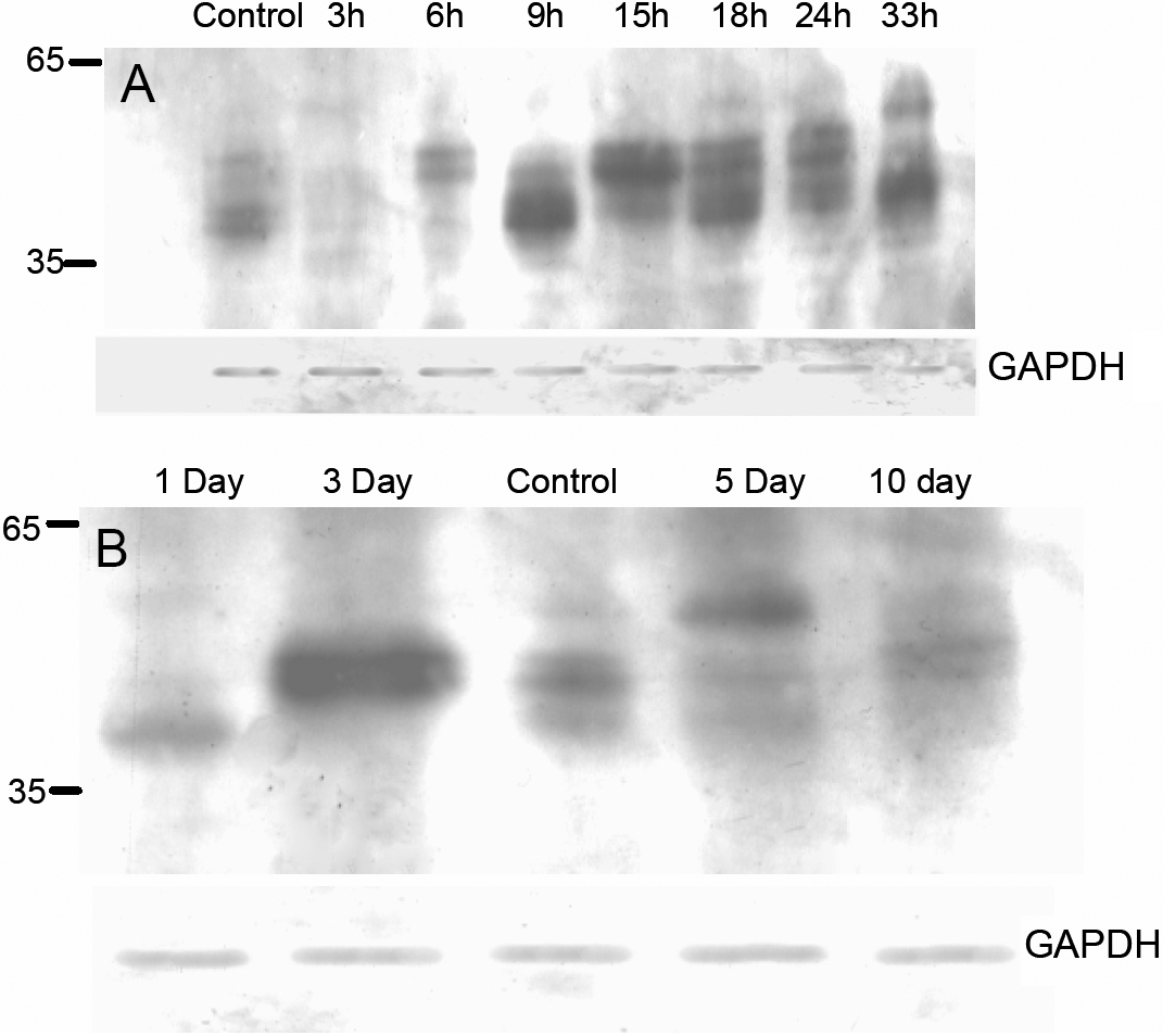
 Figure 5 of Govindarajan, Mol Vis 2009; 15:1079-1091.