Figure 7. Evaluation of biocompatibility
of the passage 191 NRCE cells on denuded amnion. A: NRCE cells
grew into a confluent cell sheet on denuded amnion 90 h later. Scale
bar: 40μm. B: SEM image showing the cell morphology of a NRCE
cell sheet on denuded amnion. Scale bar: 10 μm. C: TEM image of
a NRCE cell from a live rabbit corneal endothelium on Descement's
membrane (DM). Scale bar: 0.2 μm. D: TEM image of a NRCE cell
on denuded amnion. The tight attachment status of the NRCE cell to
denuded amnion (dAM), almost identical to that of the NRCE cell on
Descement's membrane in (C), was shown. Scale bar: 0.2 μm.
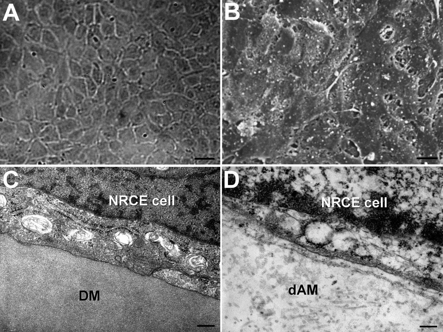
 Figure 7 of Fan, Mol Vis 2009; 15:1070-1078.