Figure 1. Morphology of NRCE cells in
vitro. A: Freshly attached NRCE cells from corneal fragments.
The non-spread polygonal cell morphology, i.e. corneal
endothelial-like, was shown. B: The monolayer formed by NRCE
cells in primary culture. The plump polygonal cell morphology was
shown. C: Passage 56 NRCE cells. The elongated polygonal cell
morphology was shown. D: Passage 227 NRCE cells. The elongated
polygonal cell morphology was shown. Scale bar: 100 μm.
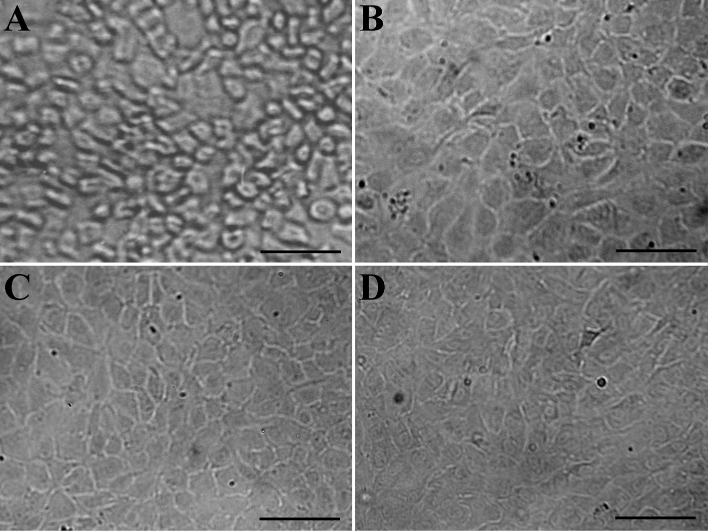
 Figure 1 of Fan, Mol Vis 2009; 15:1070-1078.