Figure 4. Expression of mucins in conjunctivas of normal mouse and transgenic mouse. A: Visualization and quantitation of conjunctival goblet cells are shown. Whole mount Alcian blue staining was used to count
the number of goblet cells in the mouse conjunctival epithelial sheets. The density of goblets cells in transgenic mice was
less than that of control mice. B: The conjunctiva of MUC5AC-DTA transgenic mouse (left column) and control littermate (right column) were subjected to mucin histological examination (100X).
In Alcian blue staining (middle panel) and mucicarmine staining (lower panel), the expression of mucin decreased in transgenic
mouse.
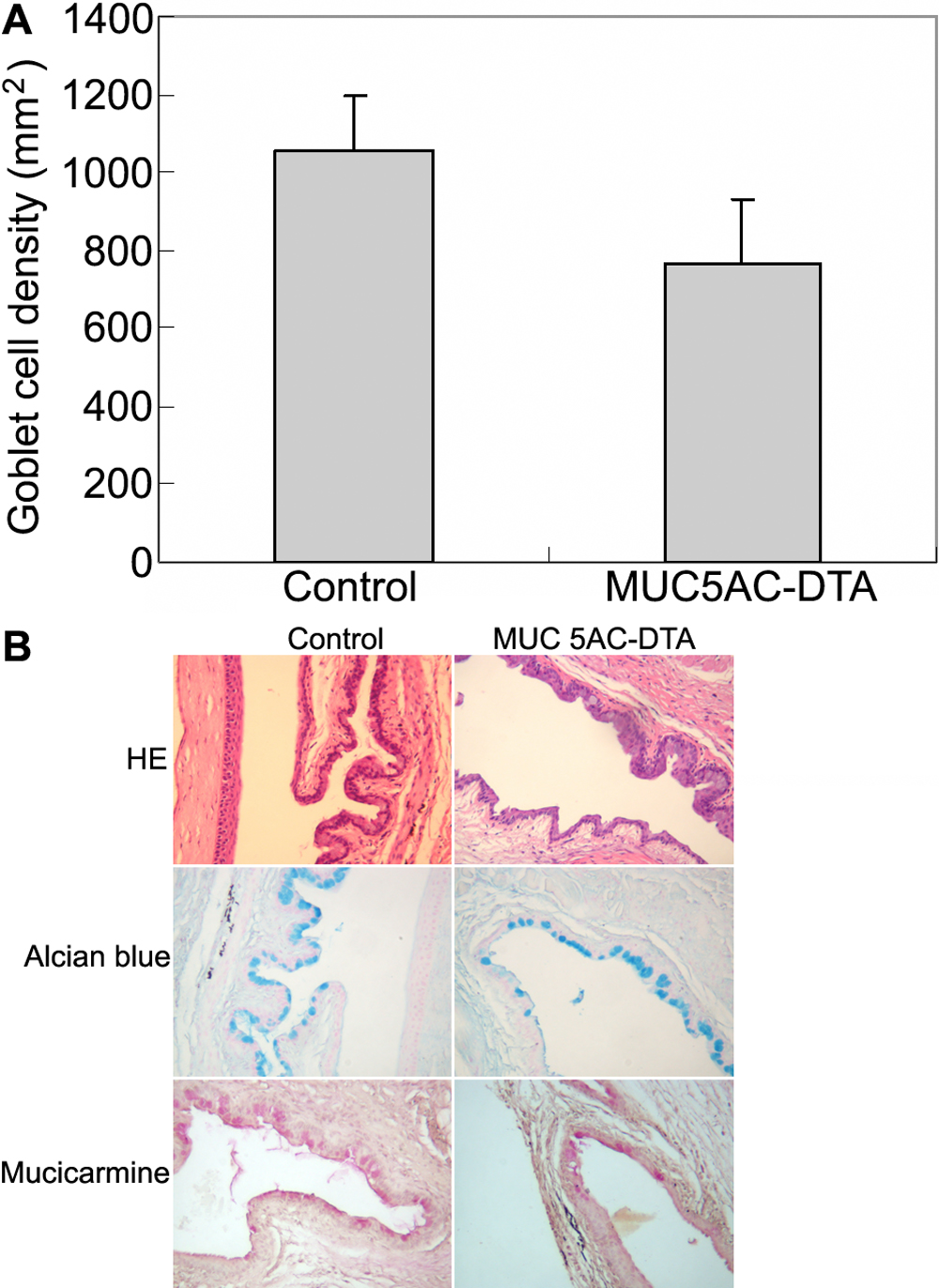
 Figure 4 of
Wang, Mol Vis 2009; 15:108-119.
Figure 4 of
Wang, Mol Vis 2009; 15:108-119.