Figure 4. Histology of burned corneas stained with hematoxylin and eosin. On day 5 (A) and 10 (B), neovascularization is observed in the limbal region of an alkali-burned cornea. Neovascularization is detected even in
the central cornea at day 20 (C,D). Bar, 50 μm.
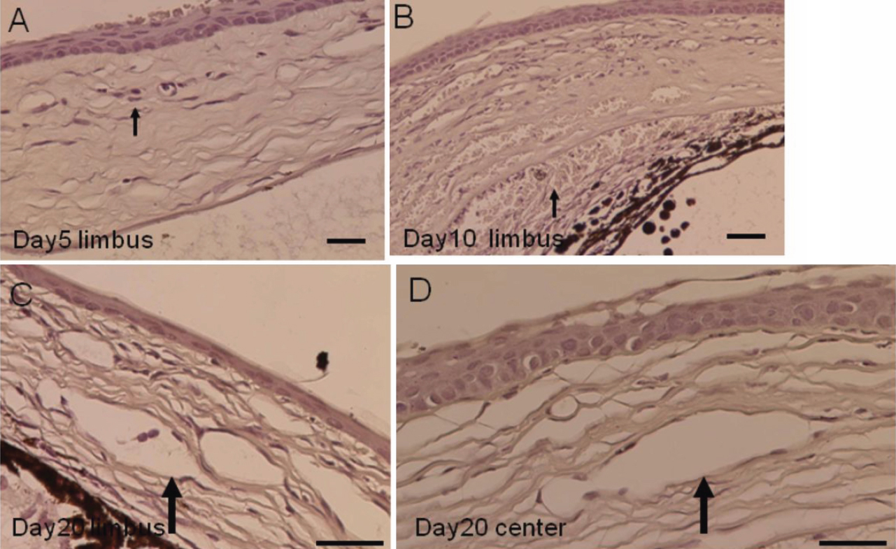
 Figure 4 of
Fujita, Mol Vis 2009; 15:1036-1044.
Figure 4 of
Fujita, Mol Vis 2009; 15:1036-1044.