Figure 3 of
Meyer, Mol Vis 2009; 15:1014-1019.
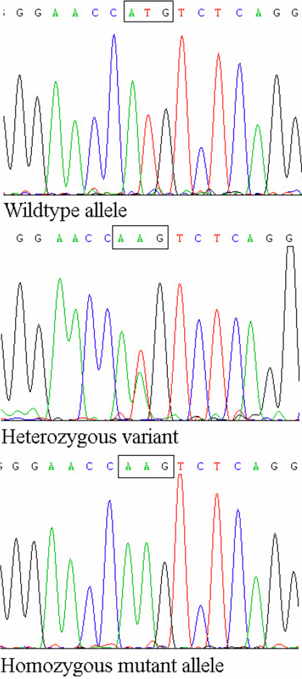
Figure 3.
CRYBB1
mutation. In top row is the wildtype sequence in a control; in the middle row is a heterozygous
CRYBB1
mutation (c.2T>A) in the mother; and at the bottom is a homozygous
CRYBB1
variant (c.2T>A) in an affected individual.
 Figure 3 of
Meyer, Mol Vis 2009; 15:1014-1019.
Figure 3 of
Meyer, Mol Vis 2009; 15:1014-1019.