Figure 6. Inhibition of the MAPK pathway decreases FGF-induced retinal regeneration in Xenopus laevis. U0126, a potent inhibitor of MEK, was used for inhibition studies at concentrations of 100 µM and 1 mM. Tadpoles were retinectomized.
Both an FGF-2-soaked heparin-coated bead and an affigel blue bead soaked in either the inhibitor or in DMSO for control were
introduced in their eyes. The pictures show representative sections of eyes collected at 30 days postsurgery and stained with
hematoxylin and eosin. A: Normal retinal regeneration was evident in the eyes that were treated with FGF-2 plus a control affigel blue bead. B: Eyes treated with FGF-2 and 100 µM U0126 affigel blue beads showed severe reduction of regeneration. C: No regeneration of the retina was detected in eyes treated with an FGF-2 bead and a 1 mM U0126 affigel blue bead. Arrows
point to regenerated neural retina. Asterisks indicate FGF-2-soaked heparin beads. Abbreviations: affigel blue bead, soaked
in the inhibitor or control (ab); cornea (C). Scale bar in C represents 100 μm and applies to all panels.
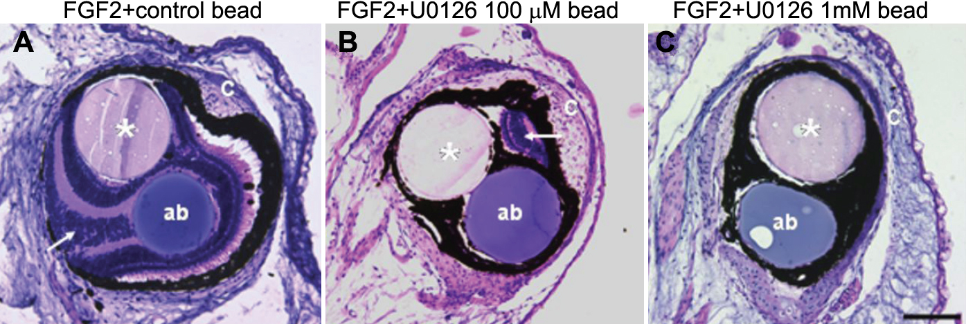
 Figure 6 of
Vergara, Mol Vis 2009; 15:1000-1013.
Figure 6 of
Vergara, Mol Vis 2009; 15:1000-1013.