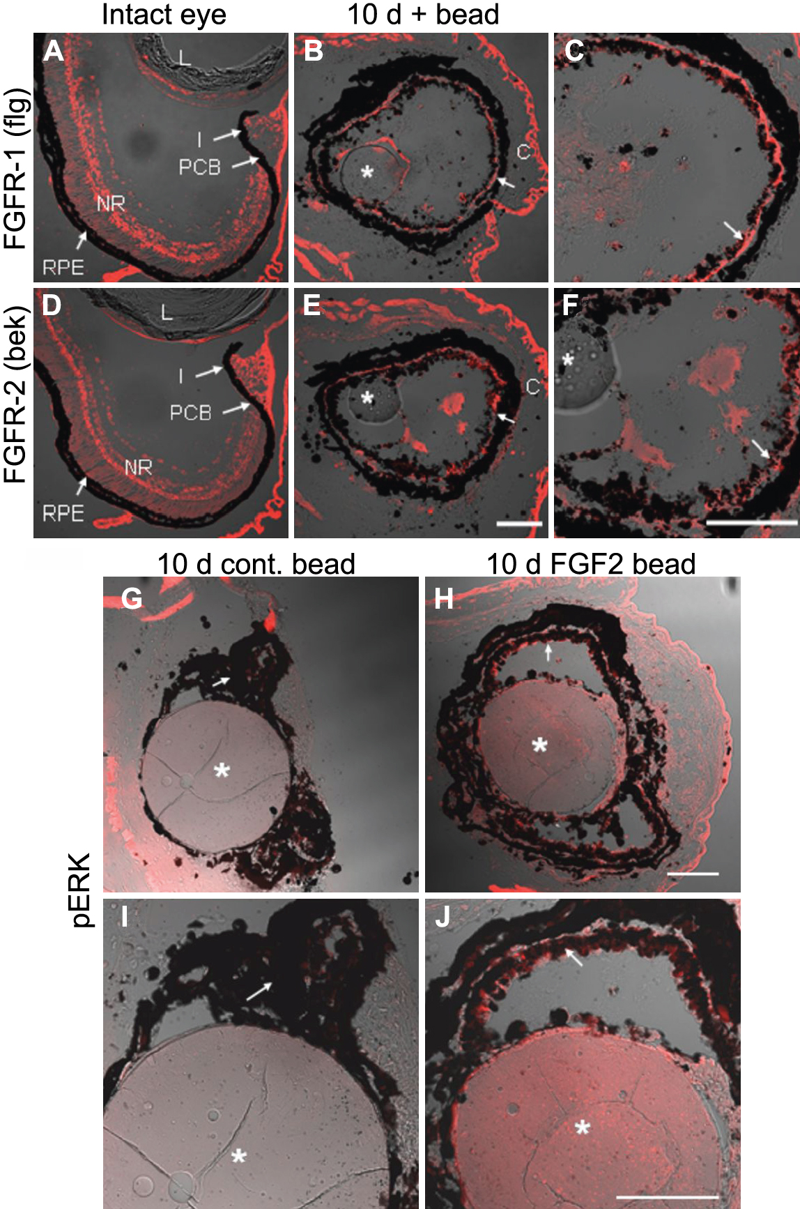
Figure 5. FGF receptors 1 and 2 expression and phosphorylated
extracellular signal-regulated protein kinase (pERK) are upregulated during regeneration.
A-
F: Immunohistochemistry for FGF receptor 1 (flg,
A-
C) and FGF receptor 2 (bek,
D-F) was performed on intact eyes (
A,
D), as well as on eyes exposed to a control bead soaked in PBS or an FGF-2 soaked bead and collected 10 days postretinectomy
(
B,
C,
E, F).
C and
F are a close up images of
B and
E respectively. Notice that FGF receptors (red) were detected in the neural retina and not in the pigmented tissues of the
intact eye, whereas after retina removal, expression of these receptors was evident in the RPE of eyes exposed to control
or FGF-2 beads (arrows).
G-J: Immunohistochemistry for pERK (red) at 10 days postretinectomy in eyes treated with control (
G, I) or FGF-2-soaked beads (
H, J).
I and
J are a close up views of
G and
H respectively. Only the pigmented epithelium of eyes exposed to FGF-2 beads was labeled by the pERK antibody. Arrows point
to the pigmented epithelium. Asterisks indicate control or FGF-2 soaked beads. Abbreviations: lens (L); neural retina (NR);
retinal pigmented epithelium (RPE); iris (I); pigmented ciliary body (PCB); cornea (C). Scale bars represent 100 μm. Scale
bar in
E applies to
A,
B,
D, and
E; scale bar in
F applies to
C and
F; scale bars in
H and
J apply to
G, H, and
I, J respectively.
 Figure 5 of
Vergara, Mol Vis 2009; 15:1000-1013.
Figure 5 of
Vergara, Mol Vis 2009; 15:1000-1013.