Figure 3. The regenerated retina is able
to form an optic nerve. Intact eyes (A, D) and eyes 30
days postretinectomy exposed to either an FGF-2 bead (B, E)
or a control bead (C, F) were injected with DiI to label
cell membranes. Ten days later, the ganglion cell axons could be seen
projecting through the optic nerve in both intact and FGF-2 exposed
eyes (D and E). Control retinectomized eyes did not
regenerate a retina and therefore did not project their axons through
what remained of the optic nerve (F). D, E, and F
correspond to fluorescent views (DiI labeling) of the bright-field
images shown in A, B, and C respectively.
Abbreviations: eyeball (E); optic nerve (ON).
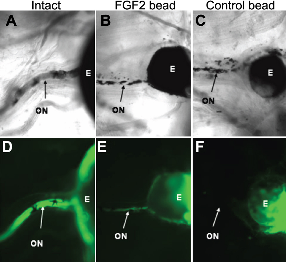
 Figure 3 of Vergara, Mol Vis 2009; 15:1000-1013.