Figure 2. Regenerated retina induced by FGF-2 expresses markers of normal retinal cells. Intact eyes (A, B, E, F, I, J, M, N, Q, R) or eyes 30 days postretinectomy with FGF-2 administration (C, D, G, H, K, L, O, P, S, T) were immunolabeled for different retinal cell markers. B, F, J, N, R, D, H, L, P, and T are close up images of the retinas shown in A, E, I, M, Q, C, G, K, O, and S, respectively. A-D: AP2α was used as a marker for amacrine cells. E-H: Islet-1 was used as a marker of ganglion cells but could also detect some subpopulations of amacrine, bipolar, and horizontal
cells. I-L: Xap-2 was used to mark rod photoreceptors. M-P: Recoverin was used to detect photoreceptors and midget cone bipolar cells. Q-T: Vimentin was used as a marker of Müller glia. Red arrows point at dark-colored Müller glia processes labeled with the vimentin
antibody. There was a general light brown background throughout the sections, whereas the staining of the antibody was actually
dark brown. All the markers tested were expressed in both the intact and regenerated retinas. Scale bars represent 100 μm
(Scale bars in O and S apply to A, E, I, M, Q, C, G, K, O, S; scale bars in P and T apply to B, F, J, N, R, D, H, L, P, T).
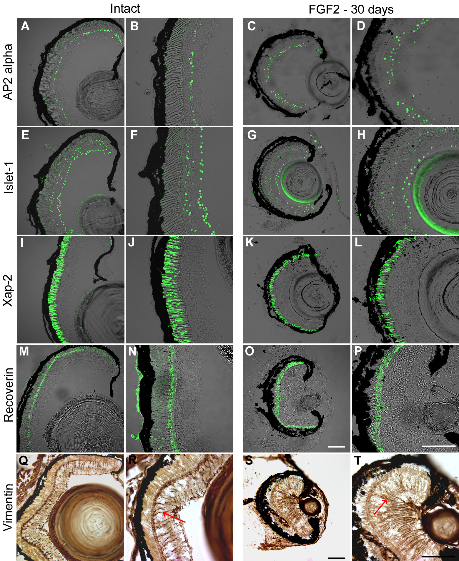
 Figure 2 of
Vergara, Mol Vis 2009; 15:1000-1013.
Figure 2 of
Vergara, Mol Vis 2009; 15:1000-1013.