Figure 1. FGF-2 induces retinal regeneration in Xenopus laevis in vivo after complete retinectomy. Shown are sections of retinectomized tadpole eyes in which a control bead (A-D) or an FGF-2-soaked bead (E-J) was introduced at day 0. The eyes were collected at postoperative days 0 (A and E), 10 (B and F), 20 (C, G, I), and 30 (D, H, J), and stained with hematoxylin and eosin. Notice the absence of nonpigmented tissues inside the eye at the earlier time points.
At 20 days, a layered neural retina was evident only in the eyes treated with FGF-2 (G); this retina was larger by 30 days (H). I and J are close up images of the regenerated retinas observed in G and H respectively. Abbreviations: optic nerve (ON); cornea (C); regenerated retina (RR); regenerated lens (RL). Asterisks indicate
control or FGF-2-soaked bead. Scale bars represent 100 μm (scale bar in H applies to A-H and scale bar in J applies also to I).
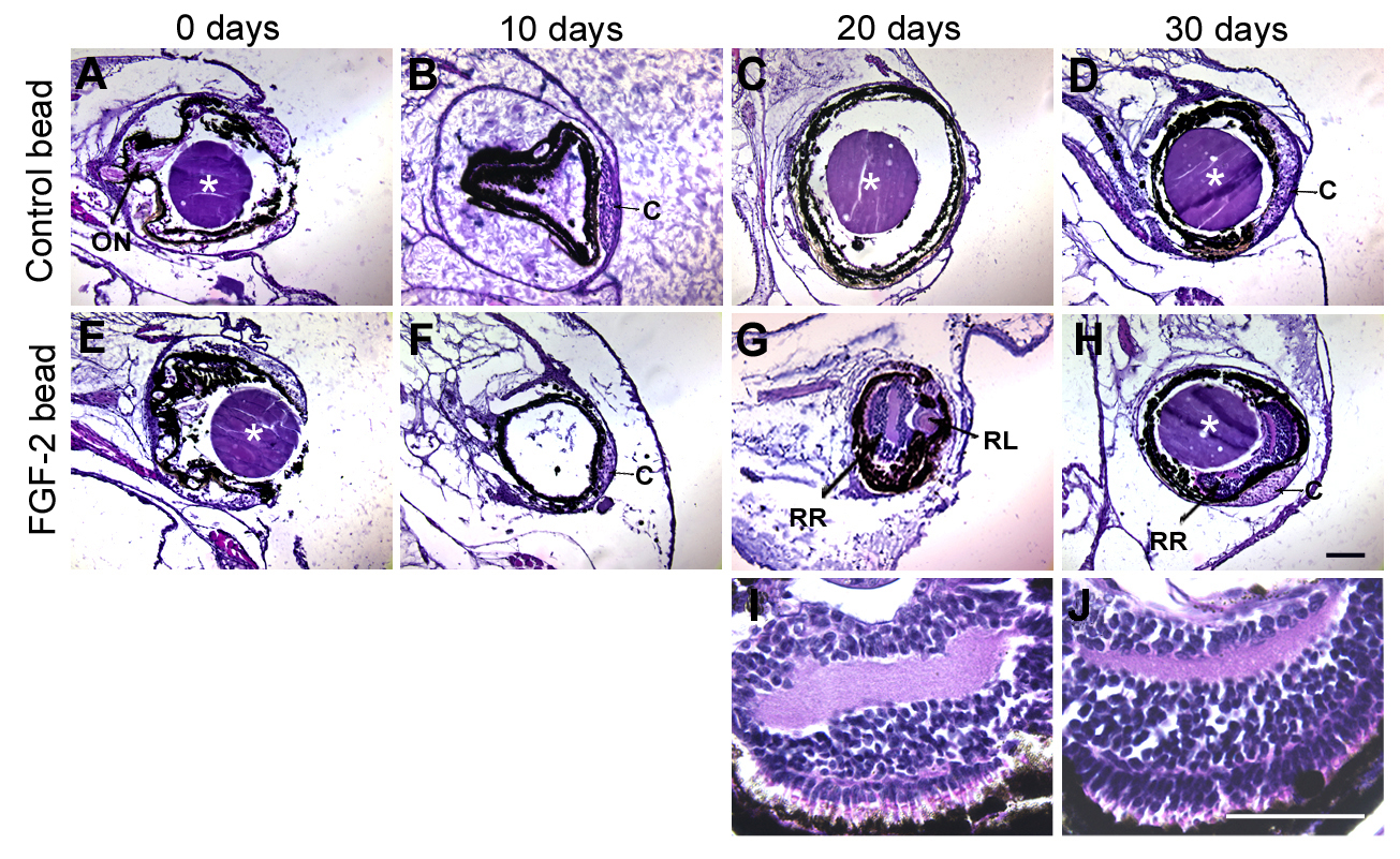
 Figure 1 of
Vergara, Mol Vis 2009; 15:1000-1013.
Figure 1 of
Vergara, Mol Vis 2009; 15:1000-1013.