Figure 3. Co-localization of MsrA and cytochrome c in lens epithelial cells. A: Co-localization of MsrA (red) and cyt c (green) and (C) merging of the two images (orange) in human lens epithelial cells
by immunofluoresence microscopy. The bottom panel shows a zoomed area of the indicated regions from the top panel. B: Co-localization of mitotracker mitochondrial marker (red) and MsrA (green) and (C) merging of the two images (orange) in
human lens epithelial cells by immunofluoresence microscopy. The bottom panel shows a zoomed area of the indicated regions
from the top panel. C: SDS-PAGE and immunoblotting of microdissected lens epithelium (E), cortex (C) and fiber (F) total protein extracts with
a cyt c-specific antibody using 100 µg of protein.
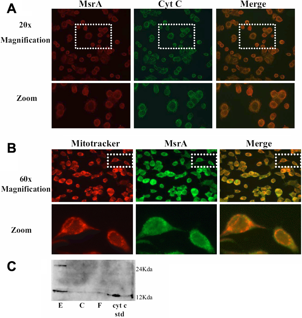
 Figure 3 of
Brennan, Mol Vis 2009; 15:985-999.
Figure 3 of
Brennan, Mol Vis 2009; 15:985-999.