Figure 1. Representative slit lamp images of the lenses of MsrA knockout mice and wild-type mice exposed to hyperbaric oxygen. A: Western blot of liver tissue supernatant (SN) and mitochondrial protein (M) isolated from wild type mice and liver and brain
tissue supernatant (SN) and mitochondrial protein (M) isolated from MsrA knockout mice using an MsrA specific antibody. B-E. Slit lamp microscopy of mouse lenses. Indicated are numbers of mice photographed, number of HBO treatments and numbers of
mice exhibiting light scattering after 8 months of treatment.
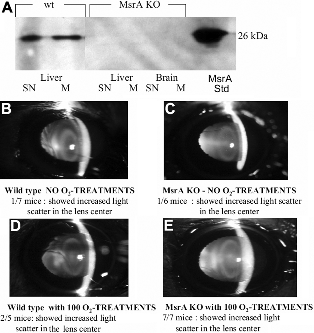
 Figure 1 of
Brennan, Mol Vis 2009; 15:985-999.
Figure 1 of
Brennan, Mol Vis 2009; 15:985-999.