Figure 5. Treated TgN S334ter rat retina photoreceptor cell nuclei counts. Animals received injections at P21 and cell counts were undertaken
at periods up to P90. Graph A represents cell nuclei counts from peripheral retina. Graph B presents cell nuclei counts from posterior pole (central) retina. Cell counts were undertaken in six eyes treated with glial-derived
neurotrophic factor (GDNF)-secreting mouse embryonic stem (mES) cells, black bars; in another 8 eyes treated with unmodified
mES cells, hatched bars; and in nine eyes treated with sham injections, white bars. Data are presented as the mean percentage
of wild-type counts±SEM and analyzed for statistical significance with a Mann–Whitney test. The asterisk indicates p<0.05.
The figure demonstrates that preservation of photoreceptor cell nuclei was seen only in eyes treated with GDNF-secreting mES
cells and mostly in peripheral retina tissue.
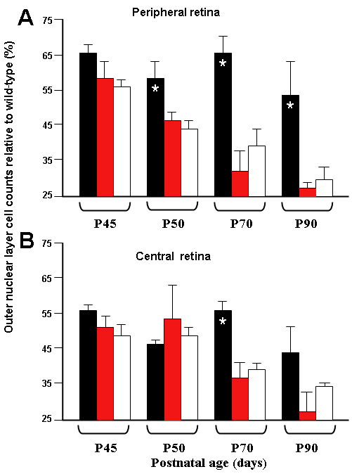
 Figure 5 of
Gregory-Evans, Mol Vis 2009; 15:962-973.
Figure 5 of
Gregory-Evans, Mol Vis 2009; 15:962-973.