Figure 4. Cryosections of TgN S334ter rat retina studied at post-natal day 35. All sections were immunostained for green fluorescent
protein (GFP; green) and retinal cell nuclei were counter-stained blue with DAPI. A-C: Illustrates images from rats from the group treated at post-natal day 21 with intravitreal injection of GDNF-secreting (and
GFP-expressing) mouse embryonic stem cells (mES cells). Plates A-C demonstrates integration of GFP-expressing mES cells into the retina. The asterisks mark clumps of mES cells. Some are seen
migrating (arrow in C) from the innermost retina toward the outer retina, and extending between the outer nuclear layer (ONL) and inner nuclear
layer (INL). D: Illustrates a mock injection eye from the group treated with intravitreal injection of sham injection of PBS, and shows autofluorescence
in the retinal pigment epithelium and photoreceptor outer segments (arrowhead). Abbreviations: inner nuclear layer (INL);
outer nuclear layer (ONL). Scale bars represent 50 μm.
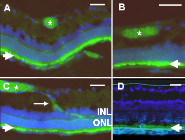
 Figure 4 of
Gregory-Evans, Mol Vis 2009; 15:962-973.
Figure 4 of
Gregory-Evans, Mol Vis 2009; 15:962-973.