Figure 3. Flatmount images of TgN S334ter rat retina treated with GDNF-secreting mES cells. Rows indicate assessments at time points
P50, P70, and P90. Columns, represent images from retina from A: upper nasal; B: upper temporal; C: central; D: lower nasal; E: lower temporal areas. All rats were from the group treated with intravitreal glial-derived neurotrophic factor (GDNF)-secreting
mouse embryonic stem (mES) cells at P21. White spots indicative of colonies of green fluorescent protein (GFP)-expressing
mES cells appear mainly on the surface of the peripheral retina rather than central retina. As time progresses, clumps become
more diffuse and ill-defined, suggesting either migration into the retinal tissue or cell loss.
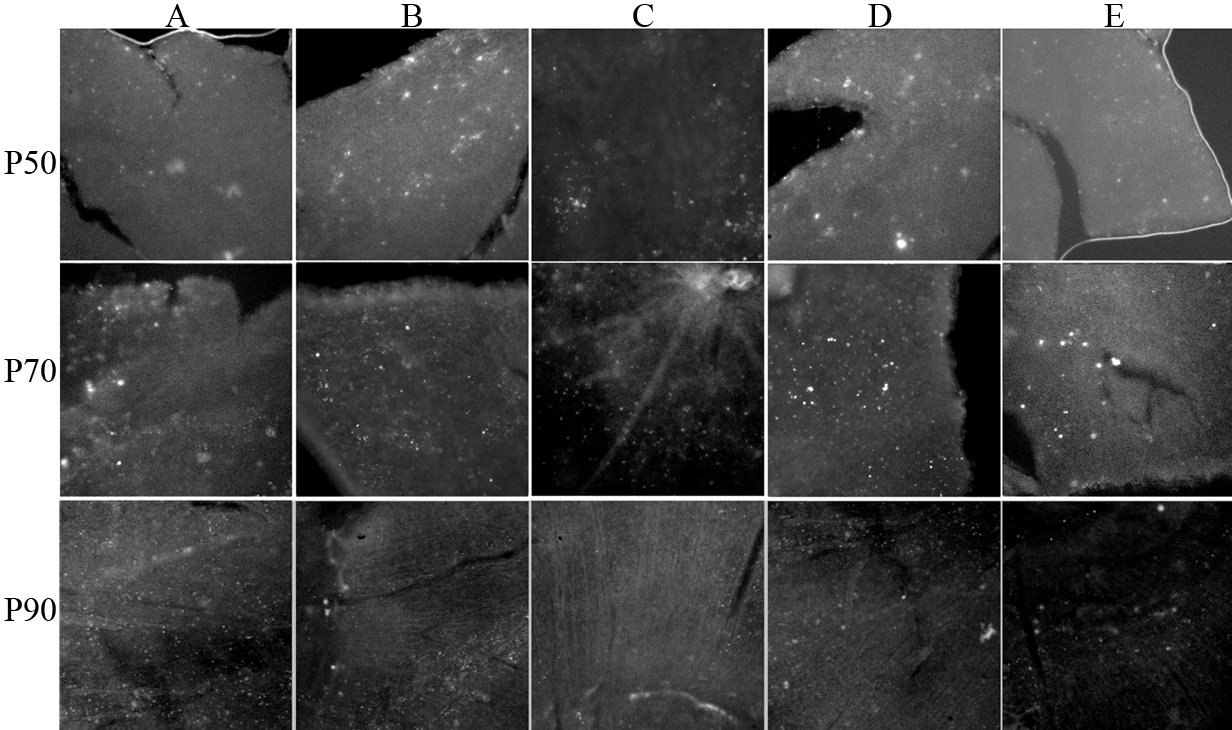
 Figure 3 of
Gregory-Evans, Mol Vis 2009; 15:962-973.
Figure 3 of
Gregory-Evans, Mol Vis 2009; 15:962-973.