Figure 2. Composite flatmount images from a single TgN S334ter treated retina at P70. One rat eye from the group treated with intravitreal
glial-derived neurotrophic factor (GDNF)-secreting mouse embryonic stem (mES) cells at P21. Spots of increased hyperfluorescence
(white), indicative of colonies of green fluorescent protein (GFP)-expressing mES cells, are mainly seen in the peripheral
retina (arrows).
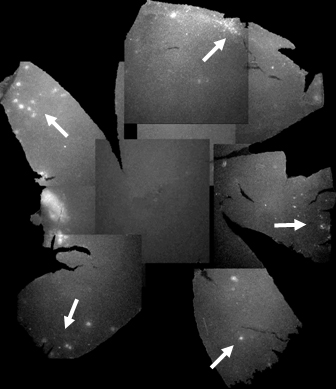
 Figure 2 of
Gregory-Evans, Mol Vis 2009; 15:962-973.
Figure 2 of
Gregory-Evans, Mol Vis 2009; 15:962-973.