Figure 1. GFP and Oct4 expression in mES cells transfected with pGDNF. Cells expressing this GDNF plasmid also express fluorescent protein
GFP and will appear red colored when exposed to GFP antibody. A: Control experiment, no red staining if mES cells are not exposed to anti-GFP antibody. B: Immunostaining of mES cells in culture using an anti-GFP antibody gives cells a red coloration confirming GFP cell expression.
C: Control experiment, no red staining if mES cells are not exposed to anti- OCT4 antibody. D: Cells immunostained using an anti-human OCT4 (POU5F1) antibody appear red, confirming Oct4 cell expression and establishing
cell totipotency.
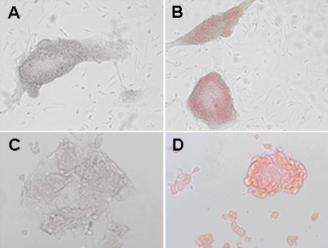
 Figure 1 of
Gregory-Evans, Mol Vis 2009; 15:962-973.
Figure 1 of
Gregory-Evans, Mol Vis 2009; 15:962-973.