Figure 4. Morphological and phenotypic of differentiated Rb-MSCs in vitro. Double staining showed expression of CK3 and BrdU both in
Group A (A-F) and Group B (H-M) as of day three. Only BrdU positive staining showed in negative controls (G,N). The red staining was CK3 and the green color was BrdU (400X).
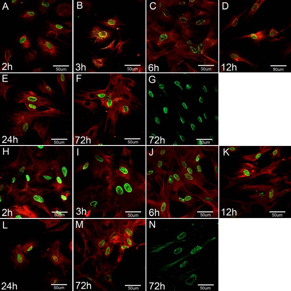
 Figure 4 of
Gu, Mol Vis 2009; 15:99-107.
Figure 4 of
Gu, Mol Vis 2009; 15:99-107.