Figure 3. Characterization of the fibrin
gel and the rabbit corneas after transplantation. Hematoxylin and eosin
staining of Group 1 (A) showed the fibrin gel with Rb-MSCs and
Group 2 (B) the fibrin gel without cells (400X). A slit-lamp
photograph (C) and hematoxylin and eosin staining (D)
showed the model of limbal stem cell deficiency (400X). A slit-lamp
photograph of the rabbit corneas after transplantation showed the
opacification and neovascularization in Group 1 (E) and in Group
2 (F). Hematoxylin and eosin staining showed that goblet cells,
new vessels, and inflammatory cells were present in some regions in
Group 1 (G) and Group 2 (H; 400X). Positive CK3 staining
was continuous throughout whole corneal epithelium in Group 1 (I),
but was irregular in Group 2 (J) by immunofluorescent staining
(100X). Double staining showed corneal epithelial cells expressed BrdU
(green) and CK3 (red) in Group 1 (K; 200X). A higher
magnification of the double staining is seen in panel L (400X).
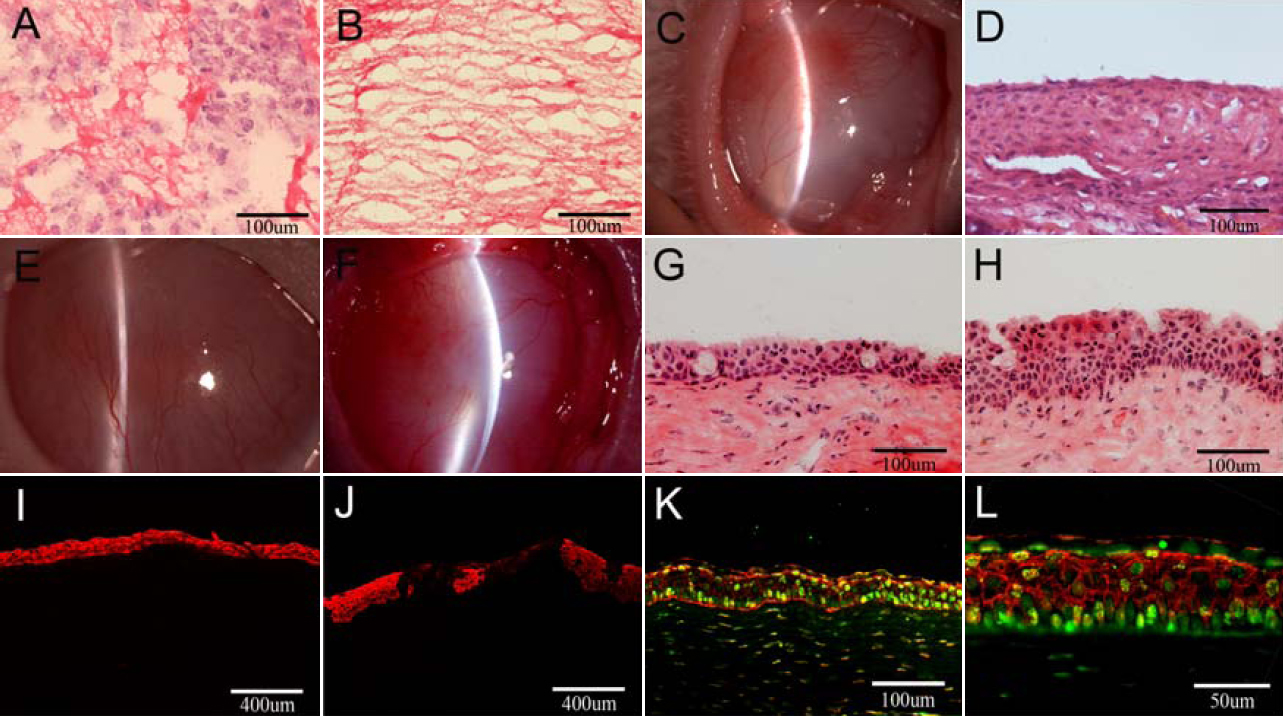
 Figure 3 of Gu, Mol Vis 2009; 15:99-107.