Figure 1. Characterization of Rb-LSCs and Rb-MSCs in vitro. A light photomicrograph of cultured Rb-LSCs is seen in panel A (200X). Positive staining of p63 (B) and integrinβ1(C) were observed on Rb-LSCs (200X). CK3 was positive on few of Rb-LSCs (D) and Hoechst 33342 (E) was used as a counterstain (200X). A light photomicrograph of cultured Rb-MSCs is seen in panel F (200×). Under the adipogenic induction medium cultured for 14 days, Rb-MSCs showed a positive reaction with oil red O stain
(G) (200X). H: Immunofluorescent staining showing Rb-MSCs labeled by BrdU expressed green fluorescence in the cell nucleus (200X).
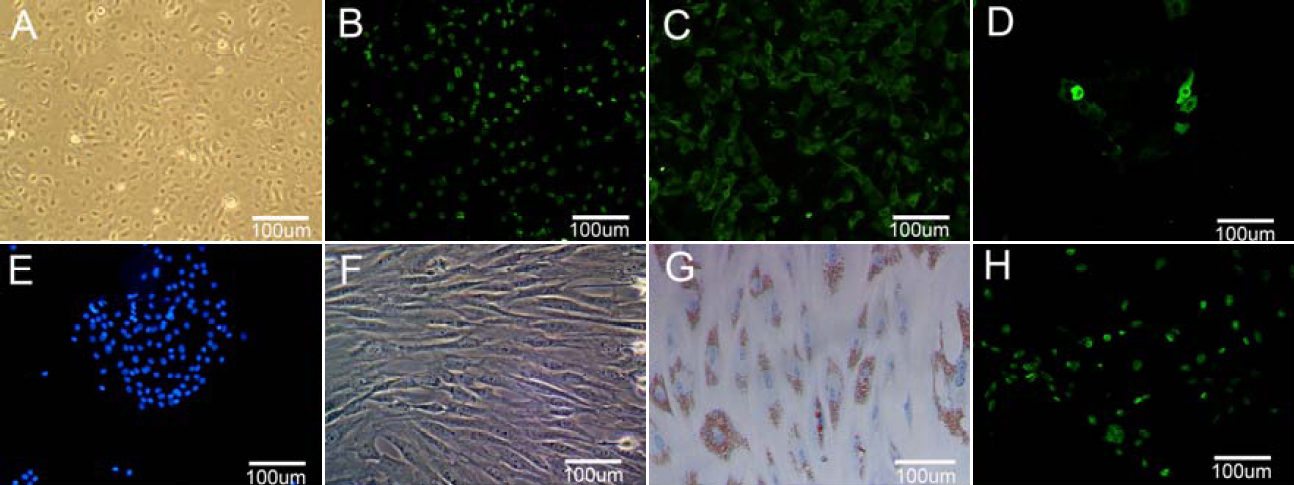
 Figure 1 of
Gu, Mol Vis 2009; 15:99-107.
Figure 1 of
Gu, Mol Vis 2009; 15:99-107.