Figure 5. Preservation of intact elongated photoreceptors in culture. Cells were dissociated using the enzymatic treatment with gentle
pipeting (Protocol A) in dark-adapted conditions. After 7 days in culture, cells were visualized under epi-fluorescence illumination
after immunostaining with anti-rhodopsin (A, D; green) and DAPI nuclear staining (B, E; blue). Merged images of rhodopsin (Rho) and DAPI (C, F, and G) showed the elongated outer segment still attached to the nucleus. Some rounded cells were still observed; but cells were,
in majority, elongated. G: Arrows indicate the outer fiber connecting the outer-inner segment to the nucleus. Scale bars represent 5 µm.
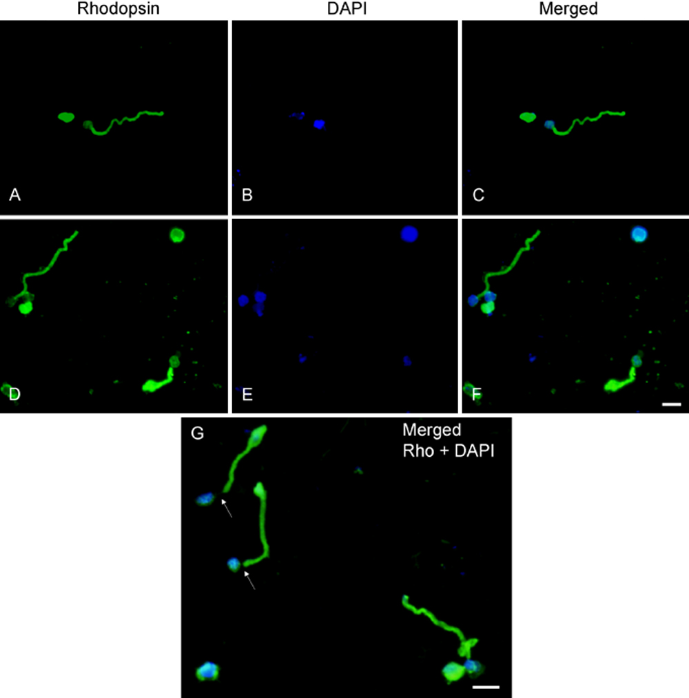
 Figure 5 of
Zayas-Santiago, Mol Vis 2009; 15:1-9.
Figure 5 of
Zayas-Santiago, Mol Vis 2009; 15:1-9.