Figure 3. Intact photoreceptors in culture
initially dissociated using three different techniques. After 2 days in
culture, cells were immunostained with anti-rhodopsin (A, E, I;
green) for outer segment labeling and with DAPI nuclear staining (B,
F, J; blue) and observed under epifluorescence illumination. A-D:
After 2 days in culture, cells dissociated using the enzymatic
treatment with gentle pipeting (Protocol A) still showed the outer
segment attached to the nucleus/cell body; however, it developed a
circular shape. E-H: In cells dissociated using the enzymatic
treatment with gentle pipeting followed by centrifugation (Protocol B)
rhodopsin was mostly expressed within the cytoplasm of the cells. I-L:
When dissociating the cells with the non-enzymatic treatment with
gentle pipeting (Protocol C) rhodopsin expression was observed in outer
segment debris. Expression of rhodopsin and DAPI within a same cell was
rare. The scale bars represent 5 μm.
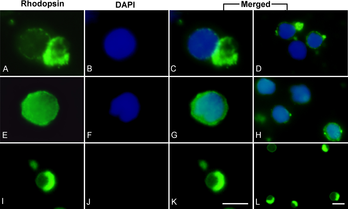
 Figure 3 of Zayas-Santiago, Mol Vis 2009; 15:1-9.