Figure 2. Quantification of intact
photoreceptors based on different dissociation techniques. Immediately
after seeded in culture, isolated intact photoreceptors were counted
under transmitted light. A: The enzymatic treatment with gentle
pipeting (Protocol A) isolated a large number of intact photoreceptors
(arrows with big arch). B: The enzymatic treatment with gentle
pipeting followed by centrifugation (Protocol B) isolated mostly cell
bodies (arrows with small arch). Most of the outer segment structures
were lost during the dissociation process. C: The non-enzymatic
treatment with gentle pipeting (Protocol C) isolated mostly elongated
outer segment structures or debris not attached to cell bodies (arrows
with dotted line). D: Shown are the percentages of intact
photoreceptors observed under transmitted light with respect to total
of observed nuclei. Protocol A yielded a significantly higher number of
intact photoreceptors compared to the other techniques (the asterisk
indicates a p<0.001). Error bars represent the standard error of the
mean. The scale bar represents 20 μm.
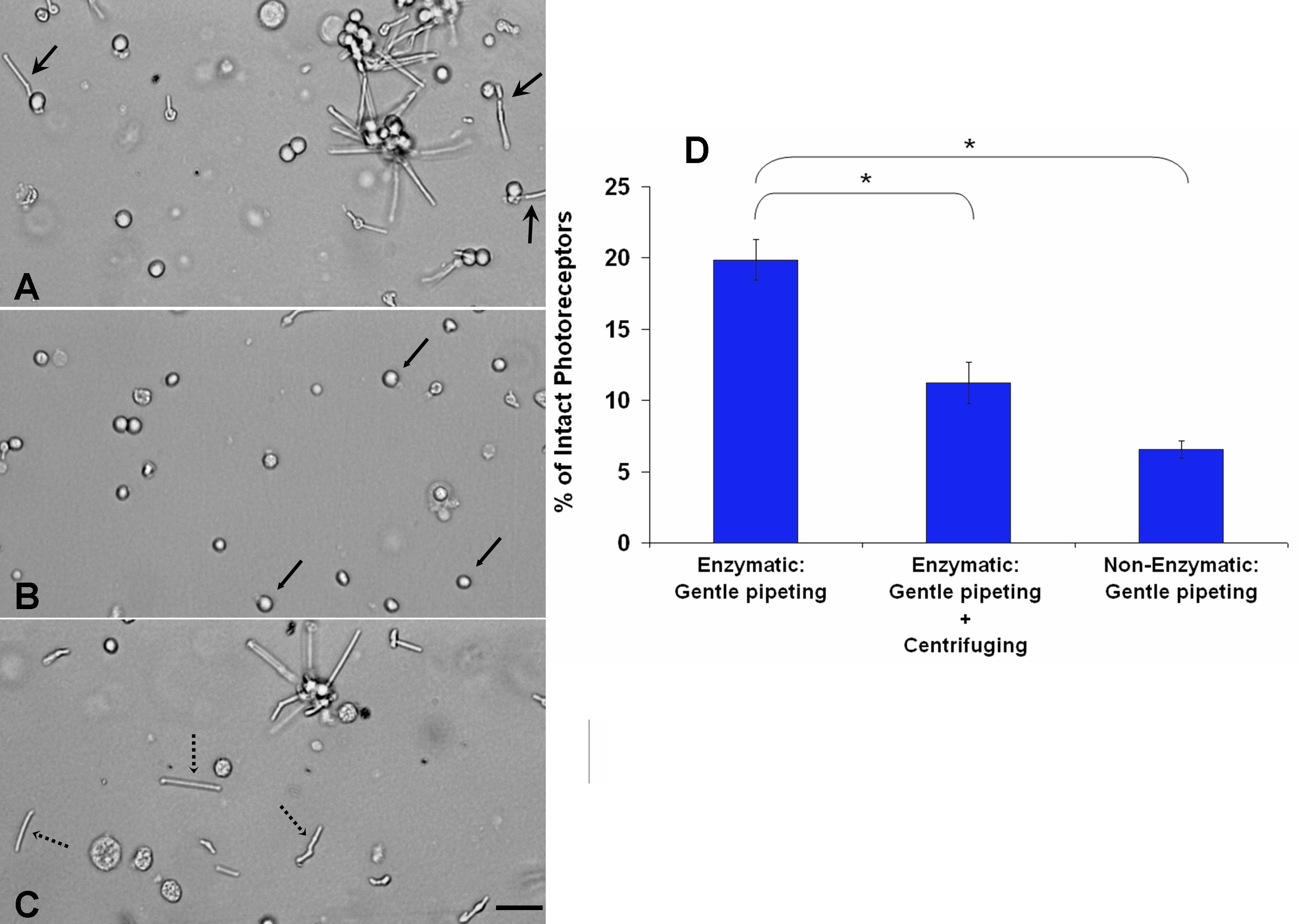
 Figure 2 of Zayas-Santiago, Mol Vis 2009; 15:1-9.