Figure 1 of
Redeker, Mol Vis 2008; 14:836-840.
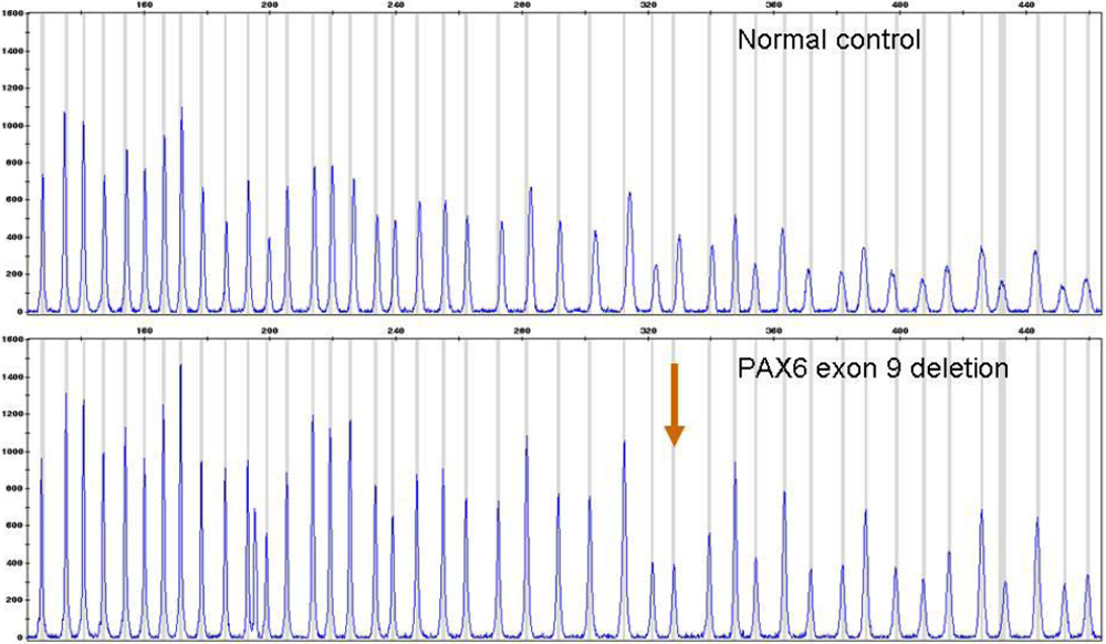
Figure 1.
Detection of
PAX6
exon 9 deletion by MLPA. Electropherograms are from a normal control and from the patient with an exon 9 deletion. The deletion is apparent by a ~50% reduction in peak area of the
PAX6
exon 9 specific probe (red arrow).