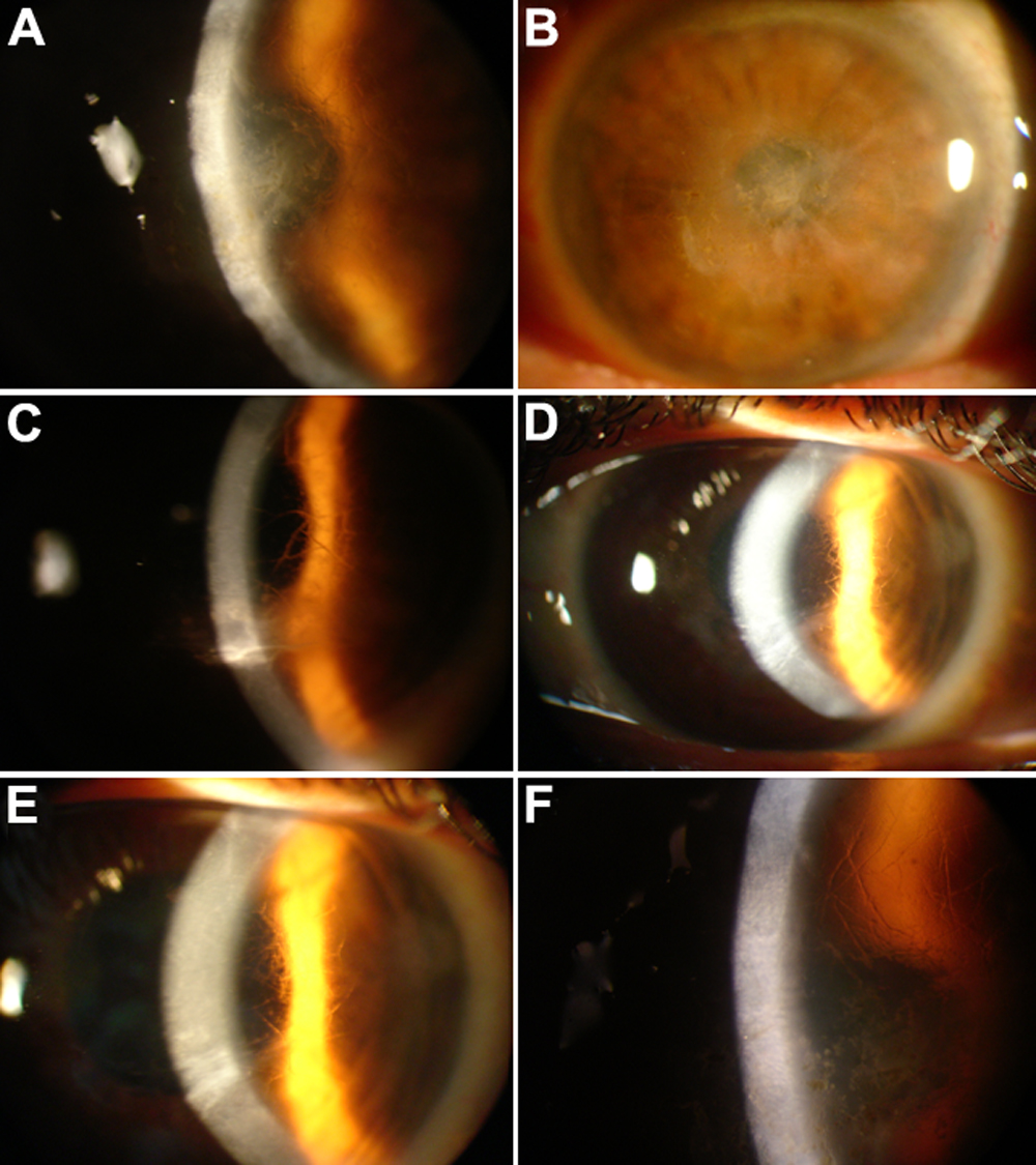
Figure 2. Corneal phenotype analyzed by
slit lamp examination. Slit lamp photographs of patient II-3 at 69
years of age show an irregularity of the epithelial surface with
subepithelial and anterior stromal scarring, resulting in diffuse
clouding of the central cornea (A and B; OD and OS,
respectively). The image of Patient III-5 at 42 years of age shows a
network of linear opacities associated with other smaller opaque spots
and refractile lattice lines (C). The photographs of case III-6
at 40 years of age show the appearance of corneal grafts (D and E).
The left cornea contains opacifications from presumed recurrent disease
(D). Fine branching lattice lines may be seen in the peripheral
anterior stroma (E). The image of patient IV-5 at eight years of
age shows the presence of large, ropy lattice lines in the anterior
stroma. A clear area is preserved around the corneoscleral limbus (F).