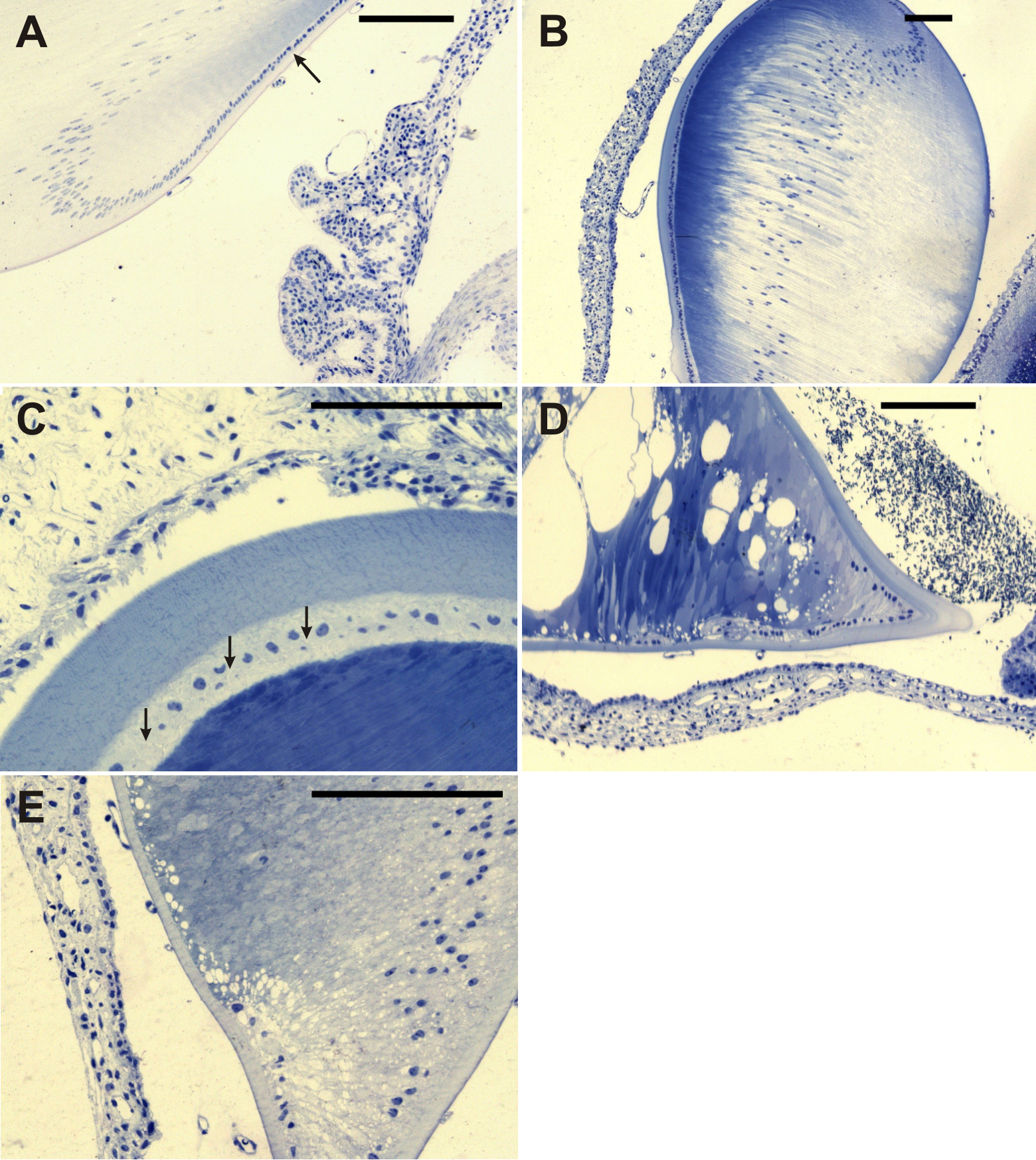
Figure 4. Histological examination of the
lens. Epon semi-thin sections were stained with toluidine blue. A:
The control animal is shown. A single layer of the cuboidal epithelium
(arrow) transforms at the equator into elongated cells representing
future lens fibers. B: The lens of +/Dca heterozygotes
corresponded in shape nearly completely with that of control animals.
At low magnification, the most pronounced feature of the lens is marked
heterogeneity in the stainability of its fibers. The lens capsule has
often variable thickness and is almost regularly thicker at the
anterior face. Persisting vascularization in the posterior eye chamber
can be observed. C: In +/Dca heterozygotes, at
the higher magnification and tangential section plane orientation,
irregularly dilated intercellular spaces between anterior cuboidal
epithelial cells (arrows) and newly formed lens fibers can be
demonstrated. D: Severe alterations of lens development were
found in homozygous Dca/Dca animals with microphthalmic eyes.
The equatorial region forms a regularly sharp angle with a distinct
thickening of the lens capsule. The anterior epithelium is
discontinuous, and sometimes there are only isolated groups of
epithelial cells visible at the anterior face of the lens. Abundant
dilatations of intercellular spaces can be seen in some lens regions as
great vacuolar formations. Also, stainability of lens fibers increases
toward the central part. E: Higher magnification of Dca/Dca
lens reveals findings similar to those in D; missing continuity
of the anterior cuboidal epithelium and striking disturbances in
organization and arrangement of cell to cell contacts.